
Contec CMS6600B Electromyogram (EMG) Machine
$1,980.00
In stock
Model NO.: CMS6600B
Channel: 4
Trademark: CONTEC
Specification: CE
HS Code: 90181990
Delivery & Availability:
Typically 5-7 working days – excluding furniture and heavy/bulky equipment. Please contact us for further information.
Description
Features:
- Professional EMG/EP operation platform and perfect test items, complete each test in the shortest time.
- Neuro and muscle navigation system, convenient in selecting test part.
- Powerful normal values system, contrast with normal data automatically.
- Flexible software design, configure system parameters according to requirements.
- High-speed data collection, electromagnetic interference suppression, Photoelectric isolation and low noise.
- Integrative hardware system, compact in configuration and easy in moving.
Performance:
- Main system part
- A/D conversion resolution 16 Bit
- Sampling ratio 200kHz
- Analysis time 5-5000ms
- Stimulating frequency 0.1-50Hz
- Amplifier part
- Four-channel
- Sensitivity 0.05μ V-20mV/Grid
- Earth noise EMG <= 4μ V(Vpp)
- EP <=0.1μ V(Vpp) (1000 times in average)
- Common-mode rejection ratio >= 100dB
- 50Hz Ban wave setting
- Upper limit for Filter-frequency 20kHz
- Lower limit for Filter-frequency 1Hz
- Gain amplifies 25 times-400000 times
- Stimulator part
- Constant current 0.2-100mA
- Pulse width 50-1000μ S
- Short circuit and overloading protection
- Auditory Stimulator
- Stimulation wave 40Hz Short, Sound Stim
- Stimulation polarity nondense wave, dense wave and alternant wave.
- Sound strength 40-120db (5db per level)
- Stimulation mode left, right, left & right
- Frequency of 40Hz carrier wave 500-8000Hz
- Visual stimulator
- Mode tessellation, horizontal bar and vertical bar.
- Stimulation view all-view, half-view and quarter-view
- Resolution 3×4, 6×8, 12×16, 24×32, 48×64
- Flash stimulator all quench, left light, right light, l&r light
Accessories:
- Disk
- Power Cord
- Power Adapter
- USB Cable
- Audio Cable
- Earth Cable
- Current Stimulator
- One-off Bipolar Needle Electrode D039035408, 0.35*40mm, D039060706, 0.60*70mm
- Reuseable Bipolar Needle Electrode R039035408, 0.35*40mm, R039060706, 0.60*70mm
- Concentric Needle Connecting Wire
- 1mm Connecting Lead Wire
- Disc Electrode
- Ring Electrode
- Surface Electrode
- Ground Electrode(Include lead cable)
- One-off Unipolar Needle Electrode
- From 1 to 2 Conversion Cable
- Surface Electrode Conversion Cable
- Fuse 0.1A/250V, 5*20mm
- Fuse F1.6AL250V, 5*20mm
- Flash Stimulator
- Audio Stimulator
Physical characteristic:
- Dimension 420mm*350mm*46mm(L× W× H)
- Net weight 6KG
Quick Comparison
| Contec CMS6600B Electromyogram (EMG) Machine remove | ASPEL AsCARD Green ECG Machine remove | Sonoscape P15 Ultrasound Machine With Four Probes remove | Sonoscape S8 Exp Portable Ultrasound remove | DrGem Diamond All-In-One Digital X-ray Machine remove | ASPEL AsCARD Grey ECG Machine remove | |
|---|---|---|---|---|---|---|
| Name | Contec CMS6600B Electromyogram (EMG) Machine remove | ASPEL AsCARD Green ECG Machine remove | Sonoscape P15 Ultrasound Machine With Four Probes remove | Sonoscape S8 Exp Portable Ultrasound remove | DrGem Diamond All-In-One Digital X-ray Machine remove | ASPEL AsCARD Grey ECG Machine remove |
| Image | 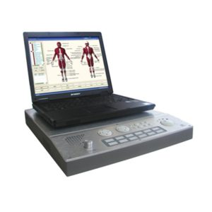 | 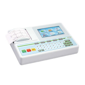 |  |  |  | 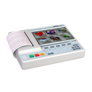 |
| SKU | SF1033560084-248 | SF1033560075-9 | SF1033560012-8 | SF1033560012-15 | SF1033560074-3 | SF1033560075-5 |
| Rating | ||||||
| Price | $1,980.00 |
| $13,900.00 | $9,350.00 |
| $1,166.00 |
| Stock | ||||||
| Availability | ||||||
| Add to cart | ||||||
| Description | In stock
Model NO.: CMS6600B
Channel: 4
Trademark: CONTEC
Specification: CE
HS Code: 90181990
| Shipped from Abroad AsCARD Green v.06.101 is a 1-, 3-, 6- and 12-channel ECG unit which enables to make electrocardiogram in full 12 leads. Intended for ECG examinations of adult and paediatric patients aimed at identification of cardiological abnormalities, myocardial ischaemia or infarction. The device is intended for use in healthcare facilities by duly trained personnel. ECG examination may be recorded in manual or automatic mode with the ability to perform the analysis and interpretation. Delivery & Availability: Typically 10 working days – excluding furniture and heavy/bulky equipment. Please contact us for further information. | In Stock A feature-rich system inheriting the Wi-Sono high-end platform, the P15 uses an array of advanced tools to help enhance the image quality. It's a cost-effective, simplified console with an intuitive user interface and multiple intelligent functions. Delivery & Availability: Typically 2 working days – excluding furniture and heavy/bulky equipment. Please contact us for further information. | Shipped from Abroad With ultra-modern innovative design and the clinically-proven technologies, S8 Exp is portable ultrasound scanner well equipped as a low-physical-effort and enhanced-image-quality ultrasound scanner, which not only provides optimized solutions for versatile applications, but does help to improve the user-experience for both routine and non-traditional challenges. Delivery & Availability: Typically 5-7 working days – excluding furniture and heavy/bulky equipment. Please contact us for further information. | Shipped from Abroad DrGem Diamond All-In-One Digital X-ray Machine is a fully automatic digital radiography system providing state-of-the-art image quality, image processing and user interface. With a wide selection of anatomical studies on the imaging software, DIAMOND automatically sets up the x-ray generator’s preprogrammed exposure technique settings, motorized radiographic stand positioning, x-ray collimation and post-image processing for the selected study. Specifically designed to increase workflow, this fully digital system offers convenient auto-positioning and advanced image processing to achieve big performance with little effort. Delivery & Availability: Typically 21 working days – excluding furniture and heavy/bulky equipment. Please contact us for further information. | Shipped from Abroad Electrocardiograph AsCARD Grey v.07.225 - is a 1, 3, 6, 12 channel ECG unit which enables to make electrocardiogram in full 12 leads. It is intended to conduct ECG examinations of adults and paediatric patients in all types of health care centres. ECG examination may be recorded in manual or automatic mode, with the possibility of analysis and interpretation. The device can be powered from 100 V ÷ 240 V mains supply or by an internal battery. Delivery & Availability: Typically 10 working days – excluding furniture and heavy/bulky equipment. Please contact us for further information. |
| Content | Features:
| AsCARD Green v.06.101 is a 1-, 3-, 6- and 12-channel ECG unit which enables to make electrocardiogram in full 12 leads. Intended for ECG examinations of adult and paediatric patients aimed at identification of cardiological abnormalities, myocardial ischaemia or infarction. The device is intended for use in healthcare facilities by duly trained personnel. ECG examination may be recorded in manual or automatic mode with the ability to perform the analysis and interpretation.
Electrocardiograph is based on advanced microprocessor technology .It is equipped with a thermal printer with high-resolution head and 4,3" LCD display. A touch panel and high-tech membrane keyboard makes this device intuitive in usage and its menu navigation exceptionally easy. This light-weight, small-footprint and battery powered cause that device can be easily transported to any location. With plastic casing and foil covered keyboard, the device is neat and easy to clean.
Technical Specifications:
Click Here To Catalogue Download | DETAILS
Super Wide-bandwidth Platform
Inheriting Wi-sono's ultra-wide system platform and with the advanced probe technology, high-resolution and deep penetration images are provided for precision medicine.
Spatial Compound Imaging
Spatial Compound Imaging utilizes several lines of sight for optimal contrast resolution, speckle reduction and border detection, with which P15 is ideal for superficial and abdominal imaging with better clarity and improved continuity of structures.
μ-Scan+
The new generation μ-Scan imaging technology gives you better image quality by reducing noise, improving signal strength and improving visualization.
Dynamic Color
Dynamic color improves upon already existing color Doppler technologies for a clearer capture of color flow and detailed visualization of even tiny veins with lower velocities.
Real-time Panoramic
With real-time panoramic, you can acquire an extended field of view for large organs or long vessels for easy measurement and diagnostic efficiency. Accomplished in real-time for the convenience of the sonographers, any mistake can also be easily back tracked and corrected without interrupting the scan.
3D/4D
Outstanding volume performance with speed and convenience makes P15 outshine others on volume imaging.
Tissue Doppler Imaging
Tissue Doppler Imaging allows clinical doctors to quantitatively evaluate local myocardial movements and functions, facilitating them with the ability to analyze and compare the motions of the different parts of the patient's heart.
Auto IMT
Quick measurement of intra-media vessel thickness ensures good reproducibility and high diagnostic efficiency.
Click Here To Download Catalogue | Sonoscape S8 Exp Portable Ultrasound scannerDETAILS Agile and Versatile With ultra-modern innovative design and the clinically-proven technologies, S8 Exp Portable Ultrasound scanner is well equipped as a low-physical-effort and enhanced-image-quality ultrasound scanner, which not only provides optimized solutions for versatile applications but does help to improve the user experience for both routine and non-traditional challenges. Working with S8 Exp, it will trigger your unlimited reverie and endow you with endless charm. Carrying forward the classical design of SonoScape's portable ultrasound products, S8 Exp successfully combines the best ergonomics, attractive design and ease of use. This charismatic identity is also enhanced by a sophisticated color palette—with sedate grey as its interior paint color and pearl white as exterior cover, S8 Exp reveals a style of aristocrat and strong character among SonoScape's ultrasound systems. Workflow The S8 Exp is a portable ultrasound scanner that adapts to your workflow, whether you are in the consulting room, at the bedside, or at a remote location. With easy-to-use new platform designed for sonographers' needs and full connection interfaces for easy connectivity and data sharing, S8 Exp leads to improved user comfort and clinical outcome as well as patient throughput and working efficiency. Powerful Platform Embedded with SonoScape's core imaging technologies such as μ-scan, PHI and Spatial Compound, S8 Exp boasts exceptional 2D image, sensitive spectral, Color and Power Doppler, displaying well-defined anatomy and pathology and facilitating a highly optimized diagnostic user environment for conclusive diagnoses. Besides, S8 Exp offers a comprehensive selection of electronic probes to maximally extend its capabilities to meet a wide range of applications including the abdomen, pediatric, OB/GYN, cardiovascular, musculoskeletal, etc. The advanced probe technologies also effectively enhance the image quality and confidence in reaching clinical diagnoses even in difficult patients.Click Here To Download Catalogue | DrGem Diamond All-In-One Digital X-ray Machine is a fully automatic digital radiography system providing state-of-the-art image quality, image processing and user interface. With a wide selection of anatomical studies on the imaging software, DIAMOND automatically sets up the x-ray generator’s pre-programmed exposure technique settings, motorized radiographic stand positioning, x-ray collimation and post-image processing for the selected study. Specifically designed to increase workflow, this fully digital system offers convenient auto-positioning and advanced image processing to achieve big performance with little effort.
Features of DrGem Diamond All-In-One Digital X-ray Machine:
Outstanding Image Quality -
Digital radiography via at panel detector improves your workflow, exam speed and comfort with efficiency. Digital at panel detector with Csl screen provides excellent spatial resolution, MTF, DQE and stability based on ne pixel pitch. A 3-field ion-chamber is provided for AEC function.
Automatic Collimation –
Automatic x-ray eld size control of the motorized collimator corresponds to dierent SIDs. Includes user adjustable lamp timer with on/oswitch.
Automatic Positioning –
Click Here To Download Catalogue |
Electrocardiograph AsCARD Grey v.07.225 - is a 1, 3, 6, 12 channel ECG unit which enables to make electrocardiogram in full 12 leads. It is intended to conduct ECG examinations of adults and paediatric patients in all types of health care centres. ECG examination may be recorded in manual or automatic mode, with the possibility of analysis and interpretation. The device can be powered from 100 V ÷ 240 V mains supply or by an internal battery.
Technical Specification:1. Visualisation of 1, 3, 6 or 12 ECG waveforms, analysis results and interpretations, examinations stored in memory.
2. Recording of 12 standard leads.
3. Print out in 1, 3, 6 or 12 ECG waveforms mode. Printing of a selected group:
Click Here To Download Catalogue |
| Weight | N/A | N/A | N/A | N/A | N/A | N/A |
| Dimensions | N/A | N/A | N/A | N/A | N/A | N/A |
| Additional information |


Reviews
There are no reviews yet.