Nio Color 2MP (MDNC‑2521)
$0.00
Shipped From Abroad
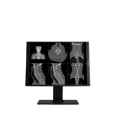
The Barco Nio Color 2MP (MDNC-2521) is a medical display designed for general radiology. With 2-megapixel resolution, DICOM-calibrated luminance, high color precision, and built-in image quality sensors, it provides dependable diagnostic viewing for everyday imaging tasks.
Typically 10-21 working days – excluding furniture and heavy/bulky equipment. Please contact us for further information.
Description
The Barco Nio Color 2MP (MDNC-2521) is a robust diagnostic display tailored for general radiology and multi-modality viewing. It offers a 1600 × 1200 resolution and a 4:3 aspect ratio, delivering precise color and grayscale rendering. Equipped with LED backlighting, 600 cd/m² DICOM-calibrated brightness, and a front sensor for image quality control, it ensures consistent performance. The unit supports high bit depth color imaging and is engineered for stable, long-term use in clinical reading rooms, PACS workstations, and diagnostic suites.
Our Nio Color 2MP display is part of a range of modern radiology monitors that give you what you need. No fluff, no overload of functionalities. But a sleek thin bezel design and just those tools and technologies that help you process cases effortlessly and efficiently.
A radiology monitor that fits your daily reading like a glove
Nio 2MP offers you exactly those tools that will make a difference. It boasts no less than 701 JNDs, so you can read detailed images with confidence. Its high luminance, I-Guard, and Uniform Luminance technologies offer you a bright and stable screen quality. And the display also contains our SteadyColor and SteadyGray technologies for stable colors and grays. In any imaging modality.
Clean desk, clear head
All of the above is built into an elegant design with thin bezels that fit both your hospital and your home office desks. Moreover, you can rotate your Nio 2MP monitor between portrait and landscape formats. Combine it in multi-head arrangements, two or three or four heads, neatly arranged in an arc embracing you.
A bright future
With Nio 2MP, you get a stable performance and a complete, industry-leading 5-year warranty that guarantees 20,000 backlight hours. Furthermore, our unique set of Intuitive Workflow Tools offers you a series of productivity hacks for more focus, flexibility, and comfort. For example, did you know that SpotView has been proven to decrease reading time by no less than 15.5%? In short, you can rest easy with this young companion. For many years to come.
Always a clear view with QAWeb Enterprise
QAWeb Enterprise helps you manage quality and assure compliance of your expanding healthcare enterprise with less effort, lower cost, and complete confidence. This fully automated and secure system supports consistent image quality, stable performance, and uptime for all PACS display systems across your enterprise. You can install QAWeb Enterprise for free on all our diagnostic and clinical review displays.
Ensuring diagnostic confidence with MDR Class IIa
Our radiology displays are MDR-certified as Class IIa. Their product information has been reviewed and cleared by independent medical and technical experts, and is audited yearly. In other words, we ensure diagnostic confidence and peace of mind for our users.
Features
-
2MP (1600 × 1200) resolution with 4:3 aspect ratio
-
DICOM-calibrated luminance (≈ 600 cd/m²)
-
High color imaging support with LED technology
-
Pixel pitch ~ 0.2707 mm
-
Front sensor (I-Guard or similar) for on-demand quality checks
-
Robust, medical-grade display for everyday radiology tasks
-
Compact form factor suitable for desktop workstations
-
Color and grayscale rendering with high fidelity
Specifications
| Category | Specification |
|---|---|
| Screen Technology | IPS-SFT Color LCD |
| Active Screen Size (Diagonal) | 541 mm (21.3″) |
| Active Screen Size (H × V) | 433 × 325 mm (17.1″ × 12.8″) |
| Aspect Ratio | 4:3 |
| Resolution | 2MP (1600 × 1200 pixels) |
| Pixel Pitch | 0.2707 mm |
| Color Imaging / Gray Imaging | Yes / Yes |
| Bit Depth | 30-bit |
| Viewing Angle (H/V) | 178° |
| Uniformity Correction | ULT |
| SteadyGray / SteadyColor | Yes (with Barco Display Controller and QAWeb Enterprise) |
| RapidFrame | Yes |
| Ambient Light Presets / Sensor | Yes / Yes |
| Backlight Output Stabilization (BLOS) | No |
| Front Sensor / Presence Sensor | Yes (I-Guard) / Yes |
| Maximum Luminance (Typical) | 1000 cd/m² |
| DICOM Calibrated Luminance | 600 cd/m² |
| Contrast Ratio | 2000:1 |
| Response Time | 12 ms (gray-to-gray average) |
| Housing Color | Black (RAL 9004) / White (RAL 9003) |
| Video Input Signals | 2 × DisplayPort 1.4 |
| Video Output Signals | N/A |
| USB Ports | 2 × USB-B 2.0 upstream; 5 × USB-A 2.0 downstream (1 charge port) |
| KVM Switch | Yes |
| Power Rating | 24 VDC, 4 A |
| Power Requirements | Adapter Technology Co., Ltd., type ATM160T-P240 <br> Input: 100–240 VAC, 1.8–0.9 A, 50–60 Hz <br> Output: 24 VDC, 6.6 A |
| Power Consumption | 37 W (nominal); <0.35 W (hibernate); <0.30 W (off) |
| Dimensions with Stand (W × H × D) | Portrait: 351 × 531~631 × 225 mm <br> Landscape: 491 × 462~562 × 225 mm |
| Dimensions without Stand (W × H × D) | Portrait: 351 × 491 × 64 mm <br> Landscape: 491 × 351 × 64 mm |
| Dimensions Packaged (W × H × D) | 455 × 210 × 770 mm |
| Net Weight with Stand | xPxx: 8.8 kg; xNxx: 7.7 kg |
| Net Weight without Stand | xPxx: 5.8 kg; xNxx: 4.7 kg |
| Net Weight Packaged | xPxx: 12.2 kg; xNxx: 11.2 kg |
| Tilt / Swivel / Pivot | -10° to +30° / ±30° / 90° |
| Height Adjustment Range | 100 mm |
| Mounting Standard | VESA (100 mm) |
| Screen Protection | xPxx: Protective, anti-reflective glass; xNxx: N/A |
| Recommended Modalities | All digital images except digital mammography |
| Certifications | CE0123, FDA 510(k) K230520, CCC, KC, BSMI, INMETRO, BIS, EAC |
| Safety Standards | IEC/EN/UL/CSA 60950-1, 62368-1, 60601-1, AAMI ES 60601-1 |
| EMI Standards | IEC/EN 60601-1-2, FCC Part 15 Class B, ICES-001 Level B, VCCI |
| Environmental Compliance | EU RoHS, China RoHS, REACH, Canada Health, WEEE, Packaging Directive |
| Supplied Accessories | User Guide, Documentation disc, System sheet, Video cables, Mains cable(s), USB cable, External power supply |
| Optional Accessories | Display controller, QA software (QAWeb Enterprise) |
| Warranty | 5 years (includes 20,000 hrs backlight warranty) |
| Operating Temperature | 0–35°C (specs: 20–30°C) |
| Storage Temperature | -20–60°C |
| Operating Humidity | 8–80% RH (non-condensing) |
| Storage Humidity | 5–85% RH (non-condensing) |
| Operating Pressure | 70 kPa |
| Storage Pressure | 50–106 kPa |
Quick Comparison
| Nio Color 2MP (MDNC‑2521) remove | Anke Supermark 1.5T MRI Machine remove | Anke Anatom 32 Fit Multi-Slice Spiral CT Scan remove | SIGNERS SUPiA X-ray Digitizer ( CR Scanner) remove | Sonoscape S8 Exp Portable Ultrasound remove | DrGem Ceiling Analogue X-ray Machine remove | ||||||||||||||||||||||||||||||||||||||||||||||||||||||||||||||||||||||||||||||||||||||||||||||||||||||||||||||||||||||||||||||||||||||||||||||||||||||||||||||||||||||||||||||||||||||||||||||||||||||||||||||||||||||||||||||||||||||||||||||||||||||||||||||||||||||||||||||||||||||||||||||||||||||||||||||||||||||||||||||||||||||||||||||||||||||||||||||||||||||||||||||||||||||||||||||||||||||||||||||||||||||||||||||||||||||||||||||||||||||||||||||||||||||||||||||||||||||||||||||||||||||||||||||||||||||||||||||||||||||||||||||||||||||||||||||||||||||||||||||||||||||||||||||||||||||||||||||||||||||||||||||||||||||||||||||||||||||||||||||||||||||||||||||||||||||||||||||||||||||||||||||||||||||||||||||||||||||||||||||||||||||||||||||||||||||||
|---|---|---|---|---|---|---|---|---|---|---|---|---|---|---|---|---|---|---|---|---|---|---|---|---|---|---|---|---|---|---|---|---|---|---|---|---|---|---|---|---|---|---|---|---|---|---|---|---|---|---|---|---|---|---|---|---|---|---|---|---|---|---|---|---|---|---|---|---|---|---|---|---|---|---|---|---|---|---|---|---|---|---|---|---|---|---|---|---|---|---|---|---|---|---|---|---|---|---|---|---|---|---|---|---|---|---|---|---|---|---|---|---|---|---|---|---|---|---|---|---|---|---|---|---|---|---|---|---|---|---|---|---|---|---|---|---|---|---|---|---|---|---|---|---|---|---|---|---|---|---|---|---|---|---|---|---|---|---|---|---|---|---|---|---|---|---|---|---|---|---|---|---|---|---|---|---|---|---|---|---|---|---|---|---|---|---|---|---|---|---|---|---|---|---|---|---|---|---|---|---|---|---|---|---|---|---|---|---|---|---|---|---|---|---|---|---|---|---|---|---|---|---|---|---|---|---|---|---|---|---|---|---|---|---|---|---|---|---|---|---|---|---|---|---|---|---|---|---|---|---|---|---|---|---|---|---|---|---|---|---|---|---|---|---|---|---|---|---|---|---|---|---|---|---|---|---|---|---|---|---|---|---|---|---|---|---|---|---|---|---|---|---|---|---|---|---|---|---|---|---|---|---|---|---|---|---|---|---|---|---|---|---|---|---|---|---|---|---|---|---|---|---|---|---|---|---|---|---|---|---|---|---|---|---|---|---|---|---|---|---|---|---|---|---|---|---|---|---|---|---|---|---|---|---|---|---|---|---|---|---|---|---|---|---|---|---|---|---|---|---|---|---|---|---|---|---|---|---|---|---|---|---|---|---|---|---|---|---|---|---|---|---|---|---|---|---|---|---|---|---|---|---|---|---|---|---|---|---|---|---|---|---|---|---|---|---|---|---|---|---|---|---|---|---|---|---|---|---|---|---|---|---|---|---|---|---|---|---|---|---|---|---|---|---|---|---|---|---|---|---|---|---|---|---|---|---|---|---|---|---|---|---|---|---|---|---|---|---|---|---|---|---|---|---|---|---|---|---|---|---|---|---|---|---|---|---|---|---|---|---|---|---|---|---|---|---|---|---|---|---|---|---|---|---|---|---|---|---|---|---|---|---|---|---|---|---|---|---|---|---|---|---|---|---|---|---|---|---|---|---|---|---|---|---|---|---|---|---|---|---|---|---|---|---|---|---|---|---|---|---|---|---|---|---|---|---|---|---|---|---|---|---|---|---|---|---|---|---|---|---|---|---|---|---|---|---|---|---|---|---|---|---|---|---|---|---|---|---|---|---|---|---|---|---|---|---|---|---|---|---|---|---|---|---|---|---|---|---|---|---|---|---|---|---|---|---|---|---|---|---|---|---|---|---|---|---|---|---|---|---|---|---|---|---|---|---|---|---|---|---|---|---|---|---|---|---|---|---|---|---|---|---|---|---|---|---|---|---|---|---|---|---|---|---|---|---|---|---|---|---|---|---|---|---|---|---|---|---|---|---|---|---|---|---|---|---|---|---|---|---|---|---|---|---|---|---|---|---|---|---|---|---|---|---|---|---|---|---|---|---|---|---|---|---|---|---|---|---|---|---|---|---|---|---|---|---|---|---|---|---|---|---|---|---|---|---|---|---|---|---|---|---|---|---|---|---|---|---|---|
| Name | Nio Color 2MP (MDNC‑2521) remove | Anke Supermark 1.5T MRI Machine remove | Anke Anatom 32 Fit Multi-Slice Spiral CT Scan remove | SIGNERS SUPiA X-ray Digitizer ( CR Scanner) remove | Sonoscape S8 Exp Portable Ultrasound remove | DrGem Ceiling Analogue X-ray Machine remove | |||||||||||||||||||||||||||||||||||||||||||||||||||||||||||||||||||||||||||||||||||||||||||||||||||||||||||||||||||||||||||||||||||||||||||||||||||||||||||||||||||||||||||||||||||||||||||||||||||||||||||||||||||||||||||||||||||||||||||||||||||||||||||||||||||||||||||||||||||||||||||||||||||||||||||||||||||||||||||||||||||||||||||||||||||||||||||||||||||||||||||||||||||||||||||||||||||||||||||||||||||||||||||||||||||||||||||||||||||||||||||||||||||||||||||||||||||||||||||||||||||||||||||||||||||||||||||||||||||||||||||||||||||||||||||||||||||||||||||||||||||||||||||||||||||||||||||||||||||||||||||||||||||||||||||||||||||||||||||||||||||||||||||||||||||||||||||||||||||||||||||||||||||||||||||||||||||||||||||||||||||||||||||||||||||||||
| Image |  |  |  |  |  |  | |||||||||||||||||||||||||||||||||||||||||||||||||||||||||||||||||||||||||||||||||||||||||||||||||||||||||||||||||||||||||||||||||||||||||||||||||||||||||||||||||||||||||||||||||||||||||||||||||||||||||||||||||||||||||||||||||||||||||||||||||||||||||||||||||||||||||||||||||||||||||||||||||||||||||||||||||||||||||||||||||||||||||||||||||||||||||||||||||||||||||||||||||||||||||||||||||||||||||||||||||||||||||||||||||||||||||||||||||||||||||||||||||||||||||||||||||||||||||||||||||||||||||||||||||||||||||||||||||||||||||||||||||||||||||||||||||||||||||||||||||||||||||||||||||||||||||||||||||||||||||||||||||||||||||||||||||||||||||||||||||||||||||||||||||||||||||||||||||||||||||||||||||||||||||||||||||||||||||||||||||||||||||||||||||||||||
| SKU | SF1033560092-4 | SF1033560092-1 | SF1033560050-01 | SF1033560012-15 | SF1033560074-7 | ||||||||||||||||||||||||||||||||||||||||||||||||||||||||||||||||||||||||||||||||||||||||||||||||||||||||||||||||||||||||||||||||||||||||||||||||||||||||||||||||||||||||||||||||||||||||||||||||||||||||||||||||||||||||||||||||||||||||||||||||||||||||||||||||||||||||||||||||||||||||||||||||||||||||||||||||||||||||||||||||||||||||||||||||||||||||||||||||||||||||||||||||||||||||||||||||||||||||||||||||||||||||||||||||||||||||||||||||||||||||||||||||||||||||||||||||||||||||||||||||||||||||||||||||||||||||||||||||||||||||||||||||||||||||||||||||||||||||||||||||||||||||||||||||||||||||||||||||||||||||||||||||||||||||||||||||||||||||||||||||||||||||||||||||||||||||||||||||||||||||||||||||||||||||||||||||||||||||||||||||||||||||||||||||||||||||
| Rating | |||||||||||||||||||||||||||||||||||||||||||||||||||||||||||||||||||||||||||||||||||||||||||||||||||||||||||||||||||||||||||||||||||||||||||||||||||||||||||||||||||||||||||||||||||||||||||||||||||||||||||||||||||||||||||||||||||||||||||||||||||||||||||||||||||||||||||||||||||||||||||||||||||||||||||||||||||||||||||||||||||||||||||||||||||||||||||||||||||||||||||||||||||||||||||||||||||||||||||||||||||||||||||||||||||||||||||||||||||||||||||||||||||||||||||||||||||||||||||||||||||||||||||||||||||||||||||||||||||||||||||||||||||||||||||||||||||||||||||||||||||||||||||||||||||||||||||||||||||||||||||||||||||||||||||||||||||||||||||||||||||||||||||||||||||||||||||||||||||||||||||||||||||||||||||||||||||||||||||||||||||||||||||||||||||||||||||||
| Price |
|
|
| $6,930.00 | $9,350.00 |
| |||||||||||||||||||||||||||||||||||||||||||||||||||||||||||||||||||||||||||||||||||||||||||||||||||||||||||||||||||||||||||||||||||||||||||||||||||||||||||||||||||||||||||||||||||||||||||||||||||||||||||||||||||||||||||||||||||||||||||||||||||||||||||||||||||||||||||||||||||||||||||||||||||||||||||||||||||||||||||||||||||||||||||||||||||||||||||||||||||||||||||||||||||||||||||||||||||||||||||||||||||||||||||||||||||||||||||||||||||||||||||||||||||||||||||||||||||||||||||||||||||||||||||||||||||||||||||||||||||||||||||||||||||||||||||||||||||||||||||||||||||||||||||||||||||||||||||||||||||||||||||||||||||||||||||||||||||||||||||||||||||||||||||||||||||||||||||||||||||||||||||||||||||||||||||||||||||||||||||||||||||||||||||||||||||||||
| Stock | |||||||||||||||||||||||||||||||||||||||||||||||||||||||||||||||||||||||||||||||||||||||||||||||||||||||||||||||||||||||||||||||||||||||||||||||||||||||||||||||||||||||||||||||||||||||||||||||||||||||||||||||||||||||||||||||||||||||||||||||||||||||||||||||||||||||||||||||||||||||||||||||||||||||||||||||||||||||||||||||||||||||||||||||||||||||||||||||||||||||||||||||||||||||||||||||||||||||||||||||||||||||||||||||||||||||||||||||||||||||||||||||||||||||||||||||||||||||||||||||||||||||||||||||||||||||||||||||||||||||||||||||||||||||||||||||||||||||||||||||||||||||||||||||||||||||||||||||||||||||||||||||||||||||||||||||||||||||||||||||||||||||||||||||||||||||||||||||||||||||||||||||||||||||||||||||||||||||||||||||||||||||||||||||||||||||||||||
| Availability | |||||||||||||||||||||||||||||||||||||||||||||||||||||||||||||||||||||||||||||||||||||||||||||||||||||||||||||||||||||||||||||||||||||||||||||||||||||||||||||||||||||||||||||||||||||||||||||||||||||||||||||||||||||||||||||||||||||||||||||||||||||||||||||||||||||||||||||||||||||||||||||||||||||||||||||||||||||||||||||||||||||||||||||||||||||||||||||||||||||||||||||||||||||||||||||||||||||||||||||||||||||||||||||||||||||||||||||||||||||||||||||||||||||||||||||||||||||||||||||||||||||||||||||||||||||||||||||||||||||||||||||||||||||||||||||||||||||||||||||||||||||||||||||||||||||||||||||||||||||||||||||||||||||||||||||||||||||||||||||||||||||||||||||||||||||||||||||||||||||||||||||||||||||||||||||||||||||||||||||||||||||||||||||||||||||||||||||
| Add to cart | |||||||||||||||||||||||||||||||||||||||||||||||||||||||||||||||||||||||||||||||||||||||||||||||||||||||||||||||||||||||||||||||||||||||||||||||||||||||||||||||||||||||||||||||||||||||||||||||||||||||||||||||||||||||||||||||||||||||||||||||||||||||||||||||||||||||||||||||||||||||||||||||||||||||||||||||||||||||||||||||||||||||||||||||||||||||||||||||||||||||||||||||||||||||||||||||||||||||||||||||||||||||||||||||||||||||||||||||||||||||||||||||||||||||||||||||||||||||||||||||||||||||||||||||||||||||||||||||||||||||||||||||||||||||||||||||||||||||||||||||||||||||||||||||||||||||||||||||||||||||||||||||||||||||||||||||||||||||||||||||||||||||||||||||||||||||||||||||||||||||||||||||||||||||||||||||||||||||||||||||||||||||||||||||||||||||||||||
| Description | Shipped From Abroad
The Barco Nio Color 2MP (MDNC-2521) is a medical display designed for general radiology. With 2-megapixel resolution, DICOM-calibrated luminance, high color precision, and built-in image quality sensors, it provides dependable diagnostic viewing for everyday imaging tasks. Delivery & Availability:
Typically 10-21 working days – excluding furniture and heavy/bulky equipment. Please contact us for further information.
| Shipped from Abroad
SuperMark 1.5T is a new generation superconducting MRI system based on years of experience in production and research. It's applicable to whole body scan, such as, nervous system, spine, joint soft tissue, pelvic and abdominal cavity, etc
Delivery & Availability: Typically 90 working days – excluding furniture and heavy/bulky equipment. Please contact us for further information. | Shipped from Abroad
This Machine gives a possibility to perform computed tomography without any problems and on high quality level. This device is used to conduct exams of internal organs and their functioning. With its help, a physician has a possibility to assess the condition of the human body as a whole.
Delivery & Availability: Typically 90 working days – excluding furniture and heavy/bulky equipment. Please contact us for further information. | Shipped from Abroad SUPiA made by Signers offers such a better clinic environment with no chemicals, ideal space, high-resolution image quality, and affordability. Delivery & Availability: Typically 14 working days – excluding furniture and heavy/bulky equipment. Please contact us for further information. | Shipped from Abroad With ultra-modern innovative design and the clinically-proven technologies, S8 Exp is portable ultrasound scanner well equipped as a low-physical-effort and enhanced-image-quality ultrasound scanner, which not only provides optimized solutions for versatile applications, but does help to improve the user-experience for both routine and non-traditional challenges. Delivery & Availability: Typically 5-7 working days – excluding furniture and heavy/bulky equipment. Please contact us for further information. | Shipped from abroad The DrGem Ceiling Analogue X-ray Machine is a diagnostic radiography system that provides reliable high quality radiographic images with a reduced dose. The reliable high-frequency x-ray generators that are known worldwide for their excellent performance, lifetime and stability. Patient tables and wall stands are also offered. Delivery & Availability: Typically 21 working days – excluding furniture and heavy/bulky equipment. Please contact us for further information. | |||||||||||||||||||||||||||||||||||||||||||||||||||||||||||||||||||||||||||||||||||||||||||||||||||||||||||||||||||||||||||||||||||||||||||||||||||||||||||||||||||||||||||||||||||||||||||||||||||||||||||||||||||||||||||||||||||||||||||||||||||||||||||||||||||||||||||||||||||||||||||||||||||||||||||||||||||||||||||||||||||||||||||||||||||||||||||||||||||||||||||||||||||||||||||||||||||||||||||||||||||||||||||||||||||||||||||||||||||||||||||||||||||||||||||||||||||||||||||||||||||||||||||||||||||||||||||||||||||||||||||||||||||||||||||||||||||||||||||||||||||||||||||||||||||||||||||||||||||||||||||||||||||||||||||||||||||||||||||||||||||||||||||||||||||||||||||||||||||||||||||||||||||||||||||||||||||||||||||||||||||||||||||||||||||||||
| Content | The Barco Nio Color 2MP (MDNC-2521) is a robust diagnostic display tailored for general radiology and multi-modality viewing. It offers a 1600 × 1200 resolution and a 4:3 aspect ratio, delivering precise color and grayscale rendering. Equipped with LED backlighting, 600 cd/m² DICOM-calibrated brightness, and a front sensor for image quality control, it ensures consistent performance. The unit supports high bit depth color imaging and is engineered for stable, long-term use in clinical reading rooms, PACS workstations, and diagnostic suites.
Our Nio Color 2MP display is part of a range of modern radiology monitors that give you what you need. No fluff, no overload of functionalities. But a sleek thin bezel design and just those tools and technologies that help you process cases effortlessly and efficiently.
A radiology monitor that fits your daily reading like a glove Nio 2MP offers you exactly those tools that will make a difference. It boasts no less than 701 JNDs, so you can read detailed images with confidence. Its high luminance, I-Guard, and Uniform Luminance technologies offer you a bright and stable screen quality. And the display also contains our SteadyColor and SteadyGray technologies for stable colors and grays. In any imaging modality. Clean desk, clear head All of the above is built into an elegant design with thin bezels that fit both your hospital and your home office desks. Moreover, you can rotate your Nio 2MP monitor between portrait and landscape formats. Combine it in multi-head arrangements, two or three or four heads, neatly arranged in an arc embracing you. A bright future With Nio 2MP, you get a stable performance and a complete, industry-leading 5-year warranty that guarantees 20,000 backlight hours. Furthermore, our unique set of Intuitive Workflow Tools offers you a series of productivity hacks for more focus, flexibility, and comfort. For example, did you know that SpotView has been proven to decrease reading time by no less than 15.5%? In short, you can rest easy with this young companion. For many years to come. Always a clear view with QAWeb Enterprise QAWeb Enterprise helps you manage quality and assure compliance of your expanding healthcare enterprise with less effort, lower cost, and complete confidence. This fully automated and secure system supports consistent image quality, stable performance, and uptime for all PACS display systems across your enterprise. You can install QAWeb Enterprise for free on all our diagnostic and clinical review displays. Ensuring diagnostic confidence with MDR Class IIa Our radiology displays are MDR-certified as Class IIa. Their product information has been reviewed and cleared by independent medical and technical experts, and is audited yearly. In other words, we ensure diagnostic confidence and peace of mind for our users. Features
Specifications
| SuperMark 1.5T is a new generation superconducting MRI system based on years of experience in production and research. It's applicable to whole body scan, such as, nervous system, spine, joint soft tissue, pelvic and abdominal cavity, etc. SuperMark 1.5T provides not only conventional pulse sequences and clinical diagnosis functions, but also provides advanced functional applications, for instance, 3D angiography and water imaging. It adopts brand new ANKE APEX operating system which ensures easy operation and fast diagnosis.
Technical Advantages:
Click Here To Download Catalogue | This Machine gives a possibility to perform computed tomography without any problems and on high quality level. This device is used to conduct exams of internal organs and their functioning. With its help, a physician has a possibility to assess the condition of the human body as a whole.
Features:
Click Here To Download Catalogue | SUPiA X-ray Digitizer made by Signers offers such a better clinic environment with no chemicals, ideal space, high-resolution image quality, and affordability
FEATURE
Rigid Type
• No damage or scratch on image plates during scanning & erasing
• Scanning & Erasing without a roller
• No cut-off image during winter and cold period
Durability
• Extremely simple structure design
• Strong aluminum base plate
• Flip covers preventing dust from inside scanner
Barcode System
• Automatically recognising cassette sizes(14x17", 10x12", 18x24cm) by barcode reader
Compact & lightweight design
Click Here To Download Catalogue | Sonoscape S8 Exp Portable Ultrasound scannerDETAILS Agile and Versatile With ultra-modern innovative design and the clinically-proven technologies, S8 Exp Portable Ultrasound scanner is well equipped as a low-physical-effort and enhanced-image-quality ultrasound scanner, which not only provides optimized solutions for versatile applications but does help to improve the user experience for both routine and non-traditional challenges. Working with S8 Exp, it will trigger your unlimited reverie and endow you with endless charm. Carrying forward the classical design of SonoScape's portable ultrasound products, S8 Exp successfully combines the best ergonomics, attractive design and ease of use. This charismatic identity is also enhanced by a sophisticated color palette—with sedate grey as its interior paint color and pearl white as exterior cover, S8 Exp reveals a style of aristocrat and strong character among SonoScape's ultrasound systems. Workflow The S8 Exp is a portable ultrasound scanner that adapts to your workflow, whether you are in the consulting room, at the bedside, or at a remote location. With easy-to-use new platform designed for sonographers' needs and full connection interfaces for easy connectivity and data sharing, S8 Exp leads to improved user comfort and clinical outcome as well as patient throughput and working efficiency. Powerful Platform Embedded with SonoScape's core imaging technologies such as μ-scan, PHI and Spatial Compound, S8 Exp boasts exceptional 2D image, sensitive spectral, Color and Power Doppler, displaying well-defined anatomy and pathology and facilitating a highly optimized diagnostic user environment for conclusive diagnoses. Besides, S8 Exp offers a comprehensive selection of electronic probes to maximally extend its capabilities to meet a wide range of applications including the abdomen, pediatric, OB/GYN, cardiovascular, musculoskeletal, etc. The advanced probe technologies also effectively enhance the image quality and confidence in reaching clinical diagnoses even in difficult patients.Click Here To Download Catalogue | DrGem Ceiling Analogue X-ray Machine is a diagnostic radiography system X-ray Machine that provides reliable high quality radiographic images with a reduced dose. The reliable high-frequency x-ray generators that are known worldwide for their excellent performance, lifetime and stability. Patient tables and wall stands are also offered.
Features of DrGem Ceiling Analogue X-ray Machine
Click Here To Download Catalogue | |||||||||||||||||||||||||||||||||||||||||||||||||||||||||||||||||||||||||||||||||||||||||||||||||||||||||||||||||||||||||||||||||||||||||||||||||||||||||||||||||||||||||||||||||||||||||||||||||||||||||||||||||||||||||||||||||||||||||||||||||||||||||||||||||||||||||||||||||||||||||||||||||||||||||||||||||||||||||||||||||||||||||||||||||||||||||||||||||||||||||||||||||||||||||||||||||||||||||||||||||||||||||||||||||||||||||||||||||||||||||||||||||||||||||||||||||||||||||||||||||||||||||||||||||||||||||||||||||||||||||||||||||||||||||||||||||||||||||||||||||||||||||||||||||||||||||||||||||||||||||||||||||||||||||||||||||||||||||||||||||||||||||||||||||||||||||||||||||||||||||||||||||||||||||||||||||||||||||||||||||||||||||||||||||||||||
| Weight | N/A | N/A | N/A | N/A | N/A | N/A | |||||||||||||||||||||||||||||||||||||||||||||||||||||||||||||||||||||||||||||||||||||||||||||||||||||||||||||||||||||||||||||||||||||||||||||||||||||||||||||||||||||||||||||||||||||||||||||||||||||||||||||||||||||||||||||||||||||||||||||||||||||||||||||||||||||||||||||||||||||||||||||||||||||||||||||||||||||||||||||||||||||||||||||||||||||||||||||||||||||||||||||||||||||||||||||||||||||||||||||||||||||||||||||||||||||||||||||||||||||||||||||||||||||||||||||||||||||||||||||||||||||||||||||||||||||||||||||||||||||||||||||||||||||||||||||||||||||||||||||||||||||||||||||||||||||||||||||||||||||||||||||||||||||||||||||||||||||||||||||||||||||||||||||||||||||||||||||||||||||||||||||||||||||||||||||||||||||||||||||||||||||||||||||||||||||||
| Dimensions | N/A | N/A | N/A | N/A | N/A | N/A | |||||||||||||||||||||||||||||||||||||||||||||||||||||||||||||||||||||||||||||||||||||||||||||||||||||||||||||||||||||||||||||||||||||||||||||||||||||||||||||||||||||||||||||||||||||||||||||||||||||||||||||||||||||||||||||||||||||||||||||||||||||||||||||||||||||||||||||||||||||||||||||||||||||||||||||||||||||||||||||||||||||||||||||||||||||||||||||||||||||||||||||||||||||||||||||||||||||||||||||||||||||||||||||||||||||||||||||||||||||||||||||||||||||||||||||||||||||||||||||||||||||||||||||||||||||||||||||||||||||||||||||||||||||||||||||||||||||||||||||||||||||||||||||||||||||||||||||||||||||||||||||||||||||||||||||||||||||||||||||||||||||||||||||||||||||||||||||||||||||||||||||||||||||||||||||||||||||||||||||||||||||||||||||||||||||||
| Additional information |
|
|


Reviews
There are no reviews yet.