| Description | Shipped From Abroad
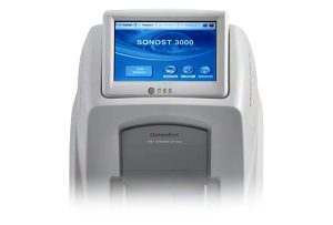
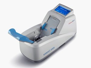
SONOST 3000 is a portable stand-alone quantitative ultrasound (QUS) bone densitometer with an embedded PC, colorful touch screen and a built-in thermal printer.
Delivery & Availability:
Typically 10-21 working days – excluding furniture and heavy/bulky equipment. Please contact us for further information.
| Shipped from Abroad
OPENMARK 5000 is 0.51T MRI. It's approved by FDA and has CE mark. It adopts two-pillar magnet design with 280 degree openness and equipped with powerful
RF and gradient system, together with advanced imaging technology, making it as a high-end system which is comparable to high-field MRI.
Delivery & Availability:
Typically 90 working days – excluding furniture and heavy/bulky equipment. Please contact us for further information. | Shipped from Abroad
The P10 color Doppler ultrasound system is a new generation product from SonoScape. It is designed to give high quality images, rich probe configurations, various clinical tools and automatic analysis software to provide you with comprehensive solutions for your growing demand for clinical applications.
Delivery & Availability:
Typically 5-7 working days – excluding furniture and heavy/bulky equipment. Please contact us for further information. | Shipped from abroad
The DrGem Ceiling Analogue X-ray Machine is a diagnostic radiography system that provides reliable high quality radiographic images with a reduced dose. The reliable high-frequency x-ray generators that are known worldwide for their excellent performance, lifetime and stability. Patient tables and wall stands are also offered.
Delivery & Availability:
Typically 21 working days – excluding furniture and heavy/bulky equipment. Please contact us for further information. | Shipped from Abroad
The ANATOM 64 CT scanner is the latest innovation for cardiac imaging based on Precision Platform system. The excellent design of Ahart technology which innovatively combined single spiral scan + gated imaging + mA modulation for easy heart imaging at extremely low radiation dose. We provide you ANATOM 64 Clarity/Precision of two models which are low/high configurations for preferences. It also offers you conventional clinical applications of low dose, better image quality and faster exams.
Delivery & Availability:
Typically 90 working days – excluding furniture and heavy/bulky equipment. Please contact us for further information. | Shipped from Abroad
SuperMark 1.5T is a new generation superconducting MRI system based on years of experience in production and research. It's applicable to whole body scan, such as, nervous system, spine, joint soft tissue, pelvic and abdominal cavity, etc
Delivery & Availability:
Typically 90 working days – excluding furniture and heavy/bulky equipment. Please contact us for further information. |
| Content | Description
https://youtu.be/bEYyQ-uEd0c?si=B6zv0KwgpYM8nTbx
The Stand-alone QUS Bone Densitometer
Portable Device with Embedded PC, Touch Screen and Thermal Printer
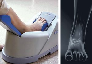
 SONOST 3000 offers a comfortable and easy-to-handle measurement through its high-sensitive touch screen without an external monitor or a keyboard. Moreover, the built-in thermal printer prints out report cards in a simple format to cut down maintenance cost. The semi-permanent waterless probe with high elasticity makes you feel comfortable on your heel bones. After the measurement, you can simply wipe out the ultrasound gel with a wet wipe. The automatic probe positioning is one of the best solutions for QUS system to minimize its positioning errors.
Features
- SOS and BUA for Measurement of BMD
The ultrasound pulse passing through the bones is significantly attenuated with diffusing signals and absorption by the spongy tissues. And QUS bone mineral density is calculated by the sound of speed (SOS) and Broadband Ultrasound Attenuation (BUA), the data obtained from a density differentiating process of sponge tissues.
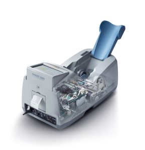
- Windows-based Embedded PC
A windows-based embedded PC enables the stand-alone mobile devices.
- USB Port and VGA Connector for External Access
SONOST 3000 can be connected to an external printer, monitor, keyboard and a mouse by USB port and VGA connector.
- Precise Result with Temperature Compensation
SONOST 3000’s algorithm for a temperature compensation function offers more precise result by taking the nearby temperature into consideration at the time of the measurement.
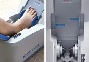
- Optimized System for Minimizing Position Errors
SONOST 3000 minimizes the possible positioning errors by offering a calf supporter, foot positioner, foot supporter, and automatic moving probe table.
- Accurate Measurement with Automatic Moving Probes
An automatic moving probe table always situates itself in the center position, so that it enhances the accuracy of a measurement.
Specifications
| Measurement method |
Ultrasound |
| Measurement site |
Calcaneus (Heel) |
| Measured parameter |
T-score, Z-score, SOS, BUA, BQI index |
| Reproducibility |
SOS – ≤ 1% CV
BUA – ≤ 2% CV |
| QC check |
Daily QC phantom |
| Dimension |
(W)615mm × (D)310mm × (H)312mm (Calf supporter folded)
(W)615mm × (D)310mm × (H)387mm |
| Weight |
12.6kg |
| OPENMARK 5000 is 0.51T MRI. It's approved by FDA and has CE mark. It adopts two-pillar magnet design with 280 degree openness and equipped with powerful
RF and gradient system, together with advanced imaging technology, making it as a high-end system which is comparable to high-field MRI.
Features:
- With the highest system stability and the highest homogeneity of the
magnet field in permanent MRI
- Screens on both sides facilitate positioning; 280 degree two-pillar magnet
design ensures stable magnet structure and facilitates interventional
treatment.
- Active and passive shimming calibrate technology ensures the magnetic
field uniformity
- Motor-driven patient couch makes it easier for patients to access and for
positioning
- Powerful hardware and software platforms ensure the scan speed, image
quality and make it possible for advanced imaging functions
- Fast scan speed eliminates motion artifact
- Rich scan sequences, advanced imaging technology and powerful postprocessing
technology ensure image quality, extend more applications,
which can fully satisfy the clinical needs
- Intelligent user-friendly operating system ensures you easy operation
Technical Specifications:
| No. |
Technique Description |
Parameter |
| 1 |
Magnet System |
|
| 1.1 |
Magnet Type |
Permanent Magnet
Automatic constant temperature
system |
| 1.2 |
Field Strength |
0.51T |
| 1.3 |
Magnet Shape |
Dual-pillar shape |
| 1.4 |
Homogeneity(40cm,DSV,VRMS) |
≤1.6ppm |
| 1.5 |
Shim Method |
Active/Passive |
| 1.6 |
Magnet Vertical Gap (Cover) |
40cm |
| 1.7 |
Magnetic Pole Dia. (Exclude Cover) |
145cm |
| 1.8 |
Accessibility(Horizontal Opening Angle, |
280° |
| 1.9 |
5 Gauss fringe field |
X-axis:horizontal ≤2.5m
Y-axis:Vertical ≤2.5m
Z-axis:horizontal ≤2.5m |
| 2 |
Patient Couch and Communication |
|
| 2.1 |
Patient Couch Driven mode |
Motor-driven |
| 2.2 |
Max. Patient Weight |
≥200kg(440lbs) |
| 2.3 |
Patient Positioning Tools |
Laser Light Localizer for positioning of center slice Motor-driven transfer to center of imaging volume |
| 2.4 |
Position accuracy |
±1mm |
| 2.5 |
Emergency Call Key |
Yes |
| 2.6 |
Intercom System |
Yes |
| 3 |
Gradient System |
|
| 3.1 |
Gradient Field Strength(Single Axis) |
≥30mT/m |
| 3.2 |
Gradient Slew Rate (Single Axis) |
≥100mT/m/ms |
| 3.3 |
Rise Time |
≤0.3ms |
| 3.4 |
Gradient Cooling System ( Gradient coils
and Power electronics) |
Air Cooling |
| 4 |
RF System |
|
| 4.1 |
RF System Type |
Digital Transmit and
Receive signal |
| 4.2 |
Number of RF Channels |
4 |
| 4.3 |
Transmitter Amplifier Peak Power |
6kW |
| 4.4 |
RF Bandwidth of Receiver |
≥1.25MHz |
| 4.5 |
Head Coil |
Yes |
| 4.6 |
Neck Coil |
Yes |
| 4.7 |
Body/Spine Coil (17 inch) |
Yes |
| 4.8 |
Body/Spine Coil (21 inch) |
Yes |
| 4.9 |
Knee Coil |
Yes |
| 4.10 |
Shoulder Coil |
Yes |
| 4.11 |
Flexible Coil |
Optional |
| 4.12 |
Breast Coil |
Optional |
| 5 |
Computer System |
|
| 5.1 |
Host Computer |
DELL Computer (for MR) |
| 5.2 |
System Software |
Windows XP |
| 5.3 |
Operation Software |
APEX |
| 5.4 |
CPU Clock rate |
3.0GHz |
| 5.5 |
Main Memory |
4GB |
| 5.6 |
Color LCD Monitor |
19” |
| 5.7 |
Keyboard and Mouse |
Standard |
| 5.8 |
Image Reconstruction Speed(256 x 256
Matrix) |
200 frame/Sec. |
| 5.9 |
Hard Disk |
500GB |
| 5.10 |
Image Storage Capacity(256 x 256
Matrix) |
500,000 |
| 5.11 |
Media Driver |
DVD RW |
| 5.12 |
DICOM 3.0 |
Yes |
| 5.13 |
Ethernet |
Yes |
| 5.14 |
Operation Console |
Yes |
| 5.15 |
Operation Chair |
Yes |
| 6 |
Scanning Parameter |
|
| 6.1 |
Max. FOV |
410mm |
| 6.2 |
Min. FOV |
5mm |
| 6.3 |
Min. TE(SE) |
5ms |
| 6.4 |
Min. TR(SE) |
11ms |
| 6.5 |
Min. TE(GR) |
1ms |
| 6.6 |
Min. TR(GR) |
3ms |
| 6.7 |
Min. 2D Thickness |
1.0mm |
| 6.8 |
Min. 3D Thickness |
0.1mm |
| 6.9 |
Max. Image Matrix |
512x512 |
| 7 |
Scanning Sequence & Imaging Technique |
|
| 7.1 |
Spin Echo 2D/3D (SE 2D/3D) |
Yes |
| 7.2 |
DE/QE |
Yes |
| 7.3 |
Fast Spin Echo 2D/3D(FSE 2D/3D) |
Yes |
| 7.4 |
Single Shot FSE 2D/3D |
Yes |
| 7.5 |
Inversion Recovery(IR) |
Yes |
| 7.6 |
Fast Inversion Recovery(FIR) |
Yes |
| 7.7 |
Gradient Echo 2D/3D(GR 2D/3D) |
Yes |
| 7.8 |
Fast GR 2D/3D |
Yes |
| 7.9 |
SPGR |
Yes |
| 7.10 |
FLAIR |
Yes |
| 7.11 |
Fat Imaging |
Yes |
| 7.12 |
Fat Suppression imaging |
Yes |
| 7.13 |
Water-Fat Separation imaging |
Yes |
| 7.14 |
TOF MRA(2D/3D) |
Yes |
| 7.15 |
MRCP(2D/3D) |
Yes |
| 7.16 |
MRU (2D/3D) |
Yes |
| 7.17 |
MRM |
Yes |
| 7.18 |
Fast Hydrograph Imaging |
Yes |
| 7.19 |
Diffusion Weighted Imaging(DWI) |
Yes |
| 7.20 |
Max. b Value |
1000s/mm2 |
| 7.21 |
Breath Hold Technology |
Yes |
| 7.22 |
Magnetization Transfer Contrast(MTC) |
Yes |
| 7.23 |
Multi-slice and Angle-free Presaturation |
Yes |
| 7.24 |
Saturation Tracking |
Yes |
| 7.25 |
Maximum Intensity Projection(MIP) |
Yes |
| 7.26 |
Multi-Angle Projection(MAP) |
Yes |
| 7.27 |
3D Reconstruction |
Yes |
| 7.28 |
Multi-planar Reconstruction(MPR) |
Yes |
| 7.29 |
Multi-Artifacts Eliminating technology |
Yes |
| 7.30 |
Checking with Part Metal Implant |
Yes |
| 7.31 |
Online Image Filtration |
Yes |
| 7.32 |
Online Post Procession |
Yes |
| 7.33 |
3D Scout |
Yes |
| 7.34 |
Scanning Protocol Preset |
Yes |
| 7.35 |
Scanning Protocol Queue Waiting |
Yes |
| 7.36 |
Advanced Image Post Processing |
Yes |
| 7.37 |
Image Fusion Technology of Vascular |
Yes |
| 7.38 |
Image Fusion Technology of Spine |
Yes |
| DETAILS
B + Compound
B + Compound utilizes several lines of sight for optimal contrast resolution, speckle reduction and border detection, with which P10 is ideal for superficial and abdominal imaging with better clarity and improved continuity of structures.
μ-Scan
The new generation μ-Scan imaging technology gives you better image quality by reducing noise, improving signal strength and improving visualization.
P10 offers a comprehensive selection of electronic probes to maximize its capabilities to meet a wide range of applications including abdomen, pediatric, OB/GYN, cardiovascular, musculoskeletal, etc. The advanced probe technologies also effectively enhance the image quality and confidence in reaching clinical diagnoses, even in difficult patients.
Convex Probe 3C-A
Ideal for an abundant of application such as abdomen, gynecology, obstetrics, urology and even abdomen biopsy.
Linear Probe L741
This linear probe is designed to satisfy vascular, breast, thyroid, and other small parts diagnosis, and its adjustable parameters could also present users a clear view of MSK and deep vessels.
Phase Array Probe 3P-A
For the purpose of adult and pediatric cardiology and emergency, the phase array probe provides elaborate presets for different exam modes, even for difficult patients.
Intracavitary Probe 6V1
Intracavitary probe could face application of gynecology, urology, prostate, and its temperature detection technology not only protects the patient but also extends the service life.
| DrGem Ceiling Analogue X-ray Machine is a diagnostic radiography system X-ray Machine that provides reliable high quality radiographic images with a reduced dose. The reliable high-frequency x-ray generators that are known worldwide for their excellent performance, lifetime and stability. Patient tables and wall stands are also offered.
Features of DrGem Ceiling Analogue X-ray Machine
- TS-CSA-A (Vertical movement, 1.6m stroke, rail length 3x4meter) including HV cable 15m
- WBS-TA: Vertical movement
- V Stroke:1,450mm in Uprigh Bucky Position,
- 1,526mm in Horizontal Bucky position.
- PBT-4 is a 4 way Floating Tabletop with. A large tabletop with extended travel enables all radiography studies with minimal patient movement. Fully fat tabletop without a frame on the edge makes cleanliness and odors free
Technical Specifications of DrGem Ceiling Analogue X-ray Machine
- Power Rating - 32KW
- Generator - GXR-32S
- Rotor - Dual Speed Starter(DSS)
- Input Power - 400/480VAC, Three phase
- Line Frequency - 50/60Hz
- X-ray tube - DXT-12M, (0.6/1.2mm, 300kHU)
- Tube Voltage - 40 to 150kV, 1kV Step
- Tube Current – 10 to 640mA
- Output - 640mA@81kV, 500mA@104kV, 400mA@130kV, 320mA@150kV
- Time Range - 1ms to 10s
- mAs Range - 0.1 to 800mAs
- Reproducibility - Coecient of Variation : kV < 0.005, Time < 0.005,mAs < 0.01
- Accuracy - kV < ±(1%+1kV), mA < ±(3%+1mA), Time <±(1%+0.5ms), mAs < ±(3%+0.1mAs)
- Linearity - Coecient of Linearity < 0.01 : CL = (X1-X2)/(X1+X2), where X is mR/mAs
- Mechanical Parts:
-TS-CSA-A (Vertical movement, 1.6m, stroke rail length 3x4meter) including HV cable 15m
- PBT-4: 4 way Floating Tabletop with Elevating Feature (66cm).
- WBS-TA: a. Vertical movement
- V Stroke:1,450mm in Upright Bucky
- Position, 1,526mm in Horizontal Bucky position.
- HVC-15: 15M HV cable
- Auto Collimator
| The ANATOM 64 CT scanner is the latest innovation for cardiac imaging based on Precision Platform system. The excellent design of Ahart technology which innovatively combined single spiral scan + gated imaging + mA modulation for easy heart imaging at extremely low radiation dose. We provide you ANATOM 64 Clarity/Precision of two models which are low/high configurations for preferences. It also offers you conventional clinical applications of low dose, better image quality and faster exams.
Features:
- Modularized OptiWave HD detector features low-cost & easy maintenance, high spatial resolution and long lifetime
- Admir3D iterative technology delivers optimal dose efficiency and noise reduction without compromising image quality
- High configurations of main components ensure the best results and maximum patient throughput
- Uniquely and creatively uses 140kV and 80kV dual energy scan mode for brain imaging on 16-slice CT to offers you extraordinary image quality both in low and high density resolutions
- AdoseTM mA modulation ensures you low dose imaging without compromising image quality particularly useful in pediatric applications
- Equipped with dedicated Abast and Amast for bone and metal artifacts
- The brilliant Ahart technology enables you to experience so easy and low-dose cardiac imaging applications
Technical Specifications:
| Model |
ANATOM 64 Precision |
ANATOM 64 Fit |
| Rack type |
Low pressure slip ring |
Low pressure slip ring |
| Scan aperture |
70cm |
70cm |
| Rack Physical inclination |
± 30 ° |
N.A |
| Rack digital inclination |
± 50 ° |
± 50 ° |
| cooling method |
Air-cooled |
Air-cooled |
| Focus to the center distance |
56 cm |
53 cm |
| |
|
|
| Maximum power (non-equivalent) |
80kW |
42kW |
| Votage (kV) |
80kV / 100kV / 120kV / 140kV |
70kV / 80kV / 100kV / 120kV /
140kV |
| |
|
|
| Heat capacity |
8MHU |
5.0MHU |
| Heat dissipation rate |
931 kHU / min |
748kHU / min |
| cooling method |
Oil cool |
Oil cool |
| Large focus size |
1.1mm × 1.2mm |
1.2mm × 1.4mm |
| Small focus size |
0.6mm × 1.2mm |
0.7mm × 0.8mm |
| Tube current range |
10-670mA |
10-350mA |
| |
|
|
| Detector type |
Optiwave detectors |
Optiwave detectors |
| Number of Z-axis |
32 |
32 |
| The width of the Z-axis |
20mm |
20mm |
| The number of elements per row |
912 |
848 |
| Total number of detectors |
29184 |
27136 |
| Acquisition mode |
64x0.625, 32x0.625,
16x0.625 |
64x0.625, 32x0.625,
16x0.625 |
| |
|
|
| Scanning range |
1800mm |
1800mm |
| Horizontal positioning accuracy |
± 0.25mm |
± 0.25mm |
| weight capacity |
205kg |
205kg |
| Minimum height |
43cm |
43cm |
| Anti - collision protection device |
Yes |
Yes |
| Foot control switch |
Yes |
Yes |
| IV rack |
Yes |
Yes |
| |
|
|
| CPU |
3.5GHz |
3.5GHz |
| RAM |
16 GB × 4 |
16 GB × 4 |
| Hard drive capacity |
1T × 2 |
1T × 2 |
| Display size |
24 inch LCD monitor |
24 inch LCD monitor |
| Display resolution |
1920 × 1200 |
1920 × 1200 |
| Computer operating system |
Windows 7 |
Windows 7 |
| Image reconstruction speed |
65 frames/ second |
65 frames/ second |
| Number of image store |
1000000 |
1000000 |
| Data external storage mode |
CD / DVD / USB |
CD / DVD / USB |
| |
|
|
| Minimum Scan Time of 360 degree |
0.39sec |
0.75sec |
| Sub-millimeter acquisition layers |
64 |
64 |
| Double sub-millimeter acquisition
layers |
64 |
64 |
| Thinnest acquisition thickness |
0.625mm |
0.625mm |
| The thinnest reconstruction
thickness |
0.3125mm |
0.625mm |
| Conventional reconstruction
thickness (mm) |
0.3125 mm, 0.625 mm, 1.25 mm, 2.5
mm, 5.0 mm, 7.5 mm, 10 mm |
0.625 mm, 1.25 mm, 2.5 mm, 5.0
mm, 7.5 mm, 10 mm |
| The reconstruction matrix |
512 x 512, 1024 x 1024 |
512 x 512, 1024 x 1024 |
| Display matrix |
1024 × 1024 |
1024 × 1024 |
| Max FOV |
52cm |
50cm |
| The maximum display field of view |
70cm |
50cm |
| Maximum scan length |
1800mm |
1800mm |
| Maximum continuous helix scan
time |
120s |
120s |
| Pitch range |
0.5-1.5 |
0.5, 1.0, 1.5 |
| |
|
|
| High contrast resolution |
21 Lp / cm @ 0% MTF |
21 Lp / cm @ 0% MTF |
| Low contrast resolution |
2mm @ 0.3% |
2mm @ 0.3% |
| Image noise |
≤ 0.25 |
≤ 0.29 |
| |
|
|
| MPR |
Yes |
Yes |
| CPR |
Yes |
Yes |
| SSD |
Yes |
Yes |
| VR |
Yes |
Yes |
| MIP |
Yes |
Yes |
| MinIP |
Yes |
Yes |
| Virtual endoscopy |
Yes |
Yes |
| CT angiography |
Yes |
Yes |
| Tissue segmentation |
Yes |
Yes |
| One-key bone removal |
Yes |
Yes |
| Automatically patient table removal |
Yes |
Yes |
| Contrast Agent Automatic Tracking
Technology- bolus tracking |
Yes |
Yes |
| Automatic linkage trigger
technology |
Yes |
Yes |
| Cine mode display |
Yes |
Yes |
| Bone artifact suppression technique |
AbastTM |
AbastTM |
| Metal artifact suppression technique |
AbastTM |
AbastTM |
| Iterative reconstruction technique |
Admir3D global iteration |
Admir3D full-domain iteration |
| Low - dose children 's scanning
technology |
Yes |
Yes |
| Low - dose lung scan |
Yes |
Yes |
| Gray matter enhancement
technology |
AccuHead |
AccuHead |
| High resolution imaging of the lung |
AccuLung |
AccuLung |
| Inner ear high resolution imaging |
AccuOtica |
AccuOtica |
| Body high resolution imaging |
AccuBody |
AccuBody |
| Bone high resolution imaging |
AccuBone |
AccuBone |
| Head dual-energy imaging |
Ahead |
Ahead |
| CT perfusion imaging |
Optional |
Optional |
| Quantitative analysis of blood
vessels |
Optional |
Optional |
| Heart coronary artery imaging |
Aheart |
N.A |
| ECG gated |
Yes |
N.A |
| Low dose cardiac scan |
Yes |
N.A |
| |
|
|
| Green energy saving technology |
AccuSaving |
AccuSaving |
| Dual-energy scan technology |
Optional |
Optional |
| SuperMark 1.5T is a new generation superconducting MRI system based on years of experience in production and research. It's applicable to whole body scan, such as, nervous system, spine, joint soft tissue, pelvic and abdominal cavity, etc. SuperMark 1.5T provides not only conventional pulse sequences and clinical diagnosis functions, but also provides advanced functional applications, for instance, 3D angiography and water imaging. It adopts brand new ANKE APEX operating system which ensures easy operation and fast diagnosis.
Technical Advantages:
- Reliable short cavity superconducting magnet system with zero liquid helium
consumption
- New generation fully digitalized and extensible multichannel spectrometer
- Powerful high efficiency and high fidelity gradient system; Multi-channel PA RF
receiving coil with intelligent identification
- English operating system and high extensible computer system
- High resolution conventional clinical images; Practical advanced functional
imaging
Superconducting MRI System:
- Highly open and humanization design -> Streamlined design
- Rich sequences and technology satisfy clinical needs -> Efficient service
Low Investment:
- High cost performance superconducting MRI system
- Zero liquid helium consumption, low running and maintenance cost
- Core technology by independent R & D supports full upgrade
- Low electric consumption
- Compact magnet design, minimum installation space: 35 square meters
High Return:
- High resolution thin slice images improve diagnosis
- Short cavity magnet design makes patients comfortable
- Fast scan speed improves work efficiency
Technical Specifications:
| No. |
Technique Description |
Parameter |
| 1 |
Magnet System |
|
| 1.1 |
Magnet Type |
Permanent Magnet
Automatic constant temperature
system |
| 1.2 |
Field Strength |
0.51T |
| 1.3 |
Magnet Shape |
Dual-pillar shape |
| 1.4 |
Homogeneity(40cm,DSV,VRMS) |
≤1.6ppm |
| 1.5 |
Shim Method |
Active/Passive |
| 1.6 |
Magnet Vertical Gap (Cover) |
40cm |
| 1.7 |
Magnetic Pole Dia. (Exclude Cover) |
145cm |
| 1.8 |
Accessibility(Horizontal Opening Angle, |
280° |
| 1.9 |
5 Gauss fringe field |
X-axis:horizontal ≤2.5m
Y-axis:Vertical ≤2.5m
Z-axis:horizontal ≤2.5m |
| 2 |
Patient Couch and Communication |
|
| 2.1 |
Patient Couch Driven mode |
Motor-driven |
| 2.2 |
Max. Patient Weight |
≥200kg(440lbs) |
| 2.3 |
Patient Positioning Tools |
Laser Light Localizer for positioning of center slice Motor-driven transfer to center of imaging volume |
| 2.4 |
Position accuracy |
±1mm |
| 2.5 |
Emergency Call Key |
Yes |
| 2.6 |
Intercom System |
Yes |
| 3 |
Gradient System |
|
| 3.1 |
Gradient Field Strength(Single Axis) |
≥30mT/m |
| 3.2 |
Gradient Slew Rate (Single Axis) |
≥100mT/m/ms |
| 3.3 |
Rise Time |
≤0.3ms |
| 3.4 |
Gradient Cooling System ( Gradient coils
and Power electronics) |
Air Cooling |
| 4 |
RF System |
|
| 4.1 |
RF System Type |
Digital Transmit and
Receive signal |
| 4.2 |
Number of RF Channels |
4 |
| 4.3 |
Transmitter Amplifier Peak Power |
6kW |
| 4.4 |
RF Bandwidth of Receiver |
≥1.25MHz |
| 4.5 |
Head Coil |
Yes |
| 4.6 |
Neck Coil |
Yes |
| 4.7 |
Body/Spine Coil (17 inch) |
Yes |
| 4.8 |
Body/Spine Coil (21 inch) |
Yes |
| 4.9 |
Knee Coil |
Yes |
| 4.10 |
Shoulder Coil |
Yes |
| 4.11 |
Flexible Coil |
Optional |
| 4.12 |
Breast Coil |
Optional |
| 5 |
Computer System |
|
| 5.1 |
Host Computer |
DELL Computer (for MR) |
| 5.2 |
System Software |
Windows XP |
| 5.3 |
Operation Software |
APEX |
| 5.4 |
CPU Clock rate |
3.0GHz |
| 5.5 |
Main Memory |
4GB |
| 5.6 |
Color LCD Monitor |
19” |
| 5.7 |
Keyboard and Mouse |
Standard |
| 5.8 |
Image Reconstruction Speed(256 x 256
Matrix) |
200 frame/Sec. |
| 5.9 |
Hard Disk |
500GB |
| 5.10 |
Image Storage Capacity(256 x 256
Matrix) |
500,000 |
| 5.11 |
Media Driver |
DVD RW |
| 5.12 |
DICOM 3.0 |
Yes |
| 5.13 |
Ethernet |
Yes |
| 5.14 |
Operation Console |
Yes |
| 5.15 |
Operation Chair |
Yes |
| 6 |
Scanning Parameter |
|
| 6.1 |
Max. FOV |
410mm |
| 6.2 |
Min. FOV |
5mm |
| 6.3 |
Min. TE(SE) |
5ms |
| 6.4 |
Min. TR(SE) |
11ms |
| 6.5 |
Min. TE(GR) |
1ms |
| 6.6 |
Min. TR(GR) |
3ms |
| 6.7 |
Min. 2D Thickness |
1.0mm |
| 6.8 |
Min. 3D Thickness |
0.1mm |
| 6.9 |
Max. Image Matrix |
512x512 |
| 7 |
Scanning Sequence & Imaging Technique |
|
| 7.1 |
Spin Echo 2D/3D (SE 2D/3D) |
Yes |
| 7.2 |
DE/QE |
Yes |
| 7.3 |
Fast Spin Echo 2D/3D(FSE 2D/3D) |
Yes |
| 7.4 |
Single Shot FSE 2D/3D |
Yes |
| 7.5 |
Inversion Recovery(IR) |
Yes |
| 7.6 |
Fast Inversion Recovery(FIR) |
Yes |
| 7.7 |
Gradient Echo 2D/3D(GR 2D/3D) |
Yes |
| 7.8 |
Fast GR 2D/3D |
Yes |
| 7.9 |
SPGR |
Yes |
| 7.10 |
FLAIR |
Yes |
| 7.11 |
Fat Imaging |
Yes |
| 7.12 |
Fat Suppression imaging |
Yes |
| 7.13 |
Water-Fat Separation imaging |
Yes |
| 7.14 |
TOF MRA(2D/3D) |
Yes |
| 7.15 |
MRCP(2D/3D) |
Yes |
| 7.16 |
MRU (2D/3D) |
Yes |
| 7.17 |
MRM |
Yes |
| 7.18 |
Fast Hydrograph Imaging |
Yes |
| 7.19 |
Diffusion Weighted Imaging(DWI) |
Yes |
| 7.20 |
Max. b Value |
1000s/mm2 |
| 7.21 |
Breath Hold Technology |
Yes |
| 7.22 |
Magnetization Transfer Contrast(MTC) |
Yes |
| 7.23 |
Multi-slice and Angle-free Presaturation |
Yes |
| 7.24 |
Saturation Tracking |
Yes |
| 7.25 |
Maximum Intensity Projection(MIP) |
Yes |
| 7.26 |
Multi-Angle Projection(MAP) |
Yes |
| 7.27 |
3D Reconstruction |
Yes |
| 7.28 |
Multi-planar Reconstruction(MPR) |
Yes |
| 7.29 |
Multi-Artifacts Eliminating technology |
Yes |
| 7.30 |
Checking with Part Metal Implant |
Yes |
| 7.31 |
Online Image Filtration |
Yes |
| 7.32 |
Online Post Procession |
Yes |
| 7.33 |
3D Scout |
Yes |
| 7.34 |
Scanning Protocol Preset |
Yes |
| 7.35 |
Scanning Protocol Queue Waiting |
Yes |
| 7.36 |
Advanced Image Post Processing |
Yes |
| 7.37 |
Image Fusion Technology of Vascular |
Yes |
| 7.38 |
Image Fusion Technology of Spine |
Yes |
|


Reviews
There are no reviews yet.