


DRGEM DR System
$0.00
Ship from abroad
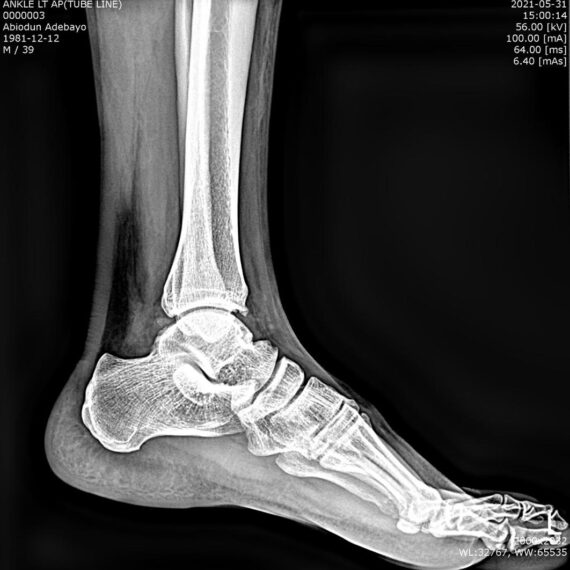
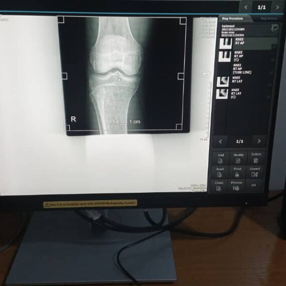
ACQUIDR is the digital imaging system composed of a Flat Panel Detector(FPD) and an imaging workstation with software. The digital FPD and full-feature imaging software with excellent digital image processing, designed for DRGEM X-ray machine.
Delivery & Availability:
Typically 21 working days – excluding furniture and heavy/bulky equipment. Please contact us for further information.

Description
DRGEM ACQUIDR (DRGEM DR System) is the digital imaging system composed of a Flat Panel Detector(FPD) and an imaging workstation with software. The digital FPD and full-feature imaging software with excellent digital image processing will meet all your needs in the diagnostic digital radiographic field.
Features of DRGEM DR System:
- Adaptable with most existing analog x-ray systems
- AED(Auto Exposure Detection) function available
- Simple and convenient installation
- Intuitive graphic user interface
- Over 1300 pre-loaded exam profiles with DRGEM’s generator
- Configurable automatic study
- PMS / EMR /HIS / RIS work list support
- Multiple printing formats
- DICOM 3.0 compliant
- Brightness and contrast adjustment
- Rotation
- Horizontal and vertical flip
- Zoom and pan
- Left / right makers
- Optimized work flow
- Automatic shutters
- Support DAP interface
- Exposure index (EI)
- DR upgrade solution by retrofit.
- Portable and Wireless FPD DR solution for maximum flexibility in virtually all general radiographic specialties.
- Portable and wireless detector fits into most existing analogue x-ray system.
- Faster workflow after DR upgrade.
- With AED(Auto Exposure Detection) function, there is no DR trigger cable between detector and generator providing very easy DR upgrade.
- Easy to interface with any kind of x-ray generator.
- Radmax acquisition workstation
- Turns any analogue X-Ray system into a fully DR system
- Optional image stitching program
- Vet software available
- Detector format: 17×17” / 17×14″, wired/wireless
Quick Comparison
| Settings | DRGEM DR System remove | SIGNERS SUPiA X-ray Digitizer ( CR Scanner) remove | Sonoscape P10 Ultrasound Machine remove | Sonoscape P50 Ultrasound Machine remove | ASPEL AsPEKT 712 Holter Monitor and Software remove | DrGem Ceiling Analogue X-ray Machine remove | ||||||||||||||||||||||||
|---|---|---|---|---|---|---|---|---|---|---|---|---|---|---|---|---|---|---|---|---|---|---|---|---|---|---|---|---|---|---|
| Name | DRGEM DR System remove | SIGNERS SUPiA X-ray Digitizer ( CR Scanner) remove | Sonoscape P10 Ultrasound Machine remove | Sonoscape P50 Ultrasound Machine remove | ASPEL AsPEKT 712 Holter Monitor and Software remove | DrGem Ceiling Analogue X-ray Machine remove | ||||||||||||||||||||||||
| Image |  |  |  |  |  |  | ||||||||||||||||||||||||
| SKU | SF1033560074-8 | SF1033560050-01 | SF1033560012-7 | SF1033560012-11 | SF1033560075-4 | SF1033560074-7 | ||||||||||||||||||||||||
| Rating | ||||||||||||||||||||||||||||||
| Price |
| $6,930.00 | $9,350.00 |
| $1,991.00 |
| ||||||||||||||||||||||||
| Stock | ||||||||||||||||||||||||||||||
| Availability | ||||||||||||||||||||||||||||||
| Add to cart | ||||||||||||||||||||||||||||||
| Description | Ship from abroad ACQUIDR is the digital imaging system composed of a Flat Panel Detector(FPD) and an imaging workstation with software. The digital FPD and full-feature imaging software with excellent digital image processing, designed for DRGEM X-ray machine. Delivery & Availability: Typically 21 working days – excluding furniture and heavy/bulky equipment. Please contact us for further information. | Shipped from Abroad SUPiA made by Signers offers such a better clinic environment with no chemicals, ideal space, high-resolution image quality, and affordability. Delivery & Availability: Typically 14 working days – excluding furniture and heavy/bulky equipment. Please contact us for further information. | Shipped from Abroad The P10 color Doppler ultrasound system is a new generation product from SonoScape. It is designed to give high quality images, rich probe configurations, various clinical tools and automatic analysis software to provide you with comprehensive solutions for your growing demand for clinical applications. Delivery & Availability: Typically 5-7 working days – excluding furniture and heavy/bulky equipment. Please contact us for further information. | Shipped from Abroad Easily accomplish more with SonoScape’s new P50 ultrasound system. Incorporating single crystal clarity, automatic corrections and calculation, and user defined flexibility promises a confident diagnostic experience as well as opening new doors of opportunity for ultrasound use. Delivery & Availability: Typically 7-14 working days – excluding furniture and heavy/bulky equipment. Please contact us for further information. | Shipped from Abroad The Holta Monitor allows quick analysis of ECG examination and detection, reviewing and editing capability in the qualitative assessment of VE, VT, Single SVE, PSVT, Pauses, Irregular Rhythm, VT, IVR, Brady - and Tachycardia, Couplets, ST-segment elevation and depression, Maximum, Minimum and averaged Heart Rates, artifacts Delivery & Availability: Typically 10 working days – excluding furniture and heavy/bulky equipment. Please contact us for further information. | Shipped from abroad The DrGem Ceiling Analogue X-ray Machine is a diagnostic radiography system that provides reliable high quality radiographic images with a reduced dose. The reliable high-frequency x-ray generators that are known worldwide for their excellent performance, lifetime and stability. Patient tables and wall stands are also offered. Delivery & Availability: Typically 21 working days – excluding furniture and heavy/bulky equipment. Please contact us for further information. | ||||||||||||||||||||||||
| Content | DRGEM ACQUIDR (DRGEM DR System) is the digital imaging system composed of a Flat Panel Detector(FPD) and an imaging workstation with software. The digital FPD and full-feature imaging software with excellent digital image processing will meet all your needs in the diagnostic digital radiographic field.
Features of DRGEM DR System:
| SUPiA X-ray Digitizer made by Signers offers such a better clinic environment with no chemicals, ideal space, high-resolution image quality, and affordability
FEATURE
Rigid Type
• No damage or scratch on image plates during scanning & erasing
• Scanning & Erasing without a roller
• No cut-off image during winter and cold period
Durability
• Extremely simple structure design
• Strong aluminum base plate
• Flip covers preventing dust from inside scanner
Barcode System
• Automatically recognising cassette sizes(14x17", 10x12", 18x24cm) by barcode reader
Compact & lightweight design
Click Here To Download Catalogue | DETAILS
B + Compound
B + Compound utilizes several lines of sight for optimal contrast resolution, speckle reduction and border detection, with which P10 is ideal for superficial and abdominal imaging with better clarity and improved continuity of structures.
μ-Scan
The new generation μ-Scan imaging technology gives you better image quality by reducing noise, improving signal strength and improving visualization.
P10 offers a comprehensive selection of electronic probes to maximize its capabilities to meet a wide range of applications including abdomen, pediatric, OB/GYN, cardiovascular, musculoskeletal, etc. The advanced probe technologies also effectively enhance the image quality and confidence in reaching clinical diagnoses, even in difficult patients.
Convex Probe 3C-A
Ideal for an abundant of application such as abdomen, gynecology, obstetrics, urology and even abdomen biopsy.
Linear Probe L741
This linear probe is designed to satisfy vascular, breast, thyroid, and other small parts diagnosis, and its adjustable parameters could also present users a clear view of MSK and deep vessels.
Phase Array Probe 3P-A
For the purpose of adult and pediatric cardiology and emergency, the phase array probe provides elaborate presets for different exam modes, even for difficult patients.
Intracavitary Probe 6V1
Intracavitary probe could face application of gynecology, urology, prostate, and its temperature detection technology not only protects the patient but also extends the service life.
Click Here To Download Catalogue | DETAILS
Powerful Compact Precision
Taking into consideration the evolving expectations and needs for ultrasound, the P50 is a slim and unobtrusive trolley system that is comfortable in tight, congested spaces with little room to work in. Providing everything you need for a comfortable examination in a small space for both you and your patient.
Single Crystal Transducer
Wideband single crystal probes greatly improve the signal ratio, acquire stunning images and provide superior sensitivity and resolution for both the near and far-fields.
μ-Scan+
The new generation μ-Scan imaging technologies give you better image quality by reducing noise, improving signal strength and improving visualization.
Dynamic Color
Dynamic colour improves upon already existing colour Doppler technologies for clear capture of colour flow and detail visualization of even tiny veins with lower velocities.
Solution for Radiology
P50, is a leading-edge ultrasound system that can meet the demands of any clinical setting. You can experience a superior performance in multi-dimensional imaging for a full range of clinical applications – abdominal, breast and cardiovascular.
C-xlasto Imaging
By understanding that tissue stiffness varies depending on the type of tissue, we can use C-xlasto Imaging to easily find abnormalities and tumours within soft tissue. The differences in tissue responses are detected and visualized in real-time by the elastography algorithms through different representations, which can be particularly helpful in analyzing breast, thyroid and musculoskeletal structures. Predominately used only in linear probes, SonoScape’s new transvaginal and bi-plane probe for gynaecology and urology are breaking the mould and expanding elastography applications.
Real-time Color Panoramic
With the combination of colour flow and real-time panoramic, visualizing the blood flow of an entire vein or artery is now an easy task. Accomplished in real-time for the convenience of the sonographers, any mistakes can also be easily backtracked and corrected without interrupting the scan.
Contrast Imaging
Contrast Imaging on P50 makes full use of the infra harmonic signal and second harmonic signal to improve the image resolution and deep penetration. What’s more, the Dynamic Acoustic Control technology effectively controls the acoustic pressure for the contrast agent, decreasing the required agent dose and assures uniform image quality, guaranteeing longer contrast agent duration and better lesion perfusion of delayed phase observation.
Solution for OB/GYN
P50 has superior image quality, automated measurement tools, and a variety of volume technologies to provide ideal solutions for clinical examinations such as pregnancy examinations, and gynecologic disease diagnosis. With a new 4D transvaginal probe, P50 helps you to see and detect fetal abnormalities and significantly improves your diagnostic confidence during your examinations.
S-Live Silhouette
A unique transparent 3D anatomical image of the fetus for improved initial anatomical review. By using this new application, the system can create completely different fetal images from conventional ultrasound images, which can depict the fetal's intracorporeal anatomical structure.
Pelvic Floor 4D
Working in conjunction with SonoScape’s latest transvaginal probes, trans-perineal 4D pelvic floor ultrasound provides a useful clinical assessment of the impact of vaginal delivery on the female anterior compartment. Allowing doctors to judge whether the pelvic organs prolapsed or not, the extent of prolapse, and determining whether the pelvic muscles tore correctly.
S-Guide
S-Guide gives the user an extensive list of example obstetric ultrasound images as reference guides and a convenient checklist system to keep track of their progress during their obstetrics examination.
Auto Face
Automatically removes masking layers in front of the fetus’s face for a clearer vision of the fetus’s face.
AVC Follicle
AVC Follicle automatically identifies how many follicles are present and calculates their individual volumes.
Solution for Cardiology
P50 provides clear 2D clinical images and Doppler sensitivity to assess critical cardiac performance. Compatible with SonoScape’s single crystal probes, the P50 can provide images with better resolution and penetration in Cardiac diagnosis.
Tissue Doppler Imaging
Tissue Doppler Imaging allows clinical doctors to quantitatively evaluate local myocardial movements and functions, facilitating them with the ability to analyze and compare the motions of the different parts of the patient’s heart.
Stress Echo
Stress echocardiography is the combination of 2D echocardiography with physical, pharmacological or electrical stress of the patient. It also then provides users with report management tools such as configurable template editor, multiple loops to select one for storage, wall motion scoring, stress echo report, etc
Auto IMT
Auto IMT is used when determining the level of vascular sclerosis present in the patient by automatically tracing and calculating the thickness of the carotid vessels. What distinguishes the P50 is that it provides an instant and accurate Mean and Max index at the touch of a single button.
Auto EF
Automated 2D Cardiac Quantification is a fully intelligent trace function for endocardium with 19 easily-adjustable points providing rapid access to proven 2D EF and volumes.
Click Here To Download Catalogue | The Holter Monitor allows quick analysis of ECG examination (arrhythmias and ST segment).
Technical specifications:
HolCARD 24W Software:
Click Here To Download Catalogue | DrGem Ceiling Analogue X-ray Machine is a diagnostic radiography system X-ray Machine that provides reliable high quality radiographic images with a reduced dose. The reliable high-frequency x-ray generators that are known worldwide for their excellent performance, lifetime and stability. Patient tables and wall stands are also offered.
Features of DrGem Ceiling Analogue X-ray Machine
Click Here To Download Catalogue | ||||||||||||||||||||||||
| Weight | N/A | N/A | N/A | N/A | N/A | N/A | ||||||||||||||||||||||||
| Dimensions | N/A | N/A | N/A | N/A | N/A | N/A | ||||||||||||||||||||||||
| Additional information |


Reviews
There are no reviews yet.