- Sorry, this product cannot be purchased.


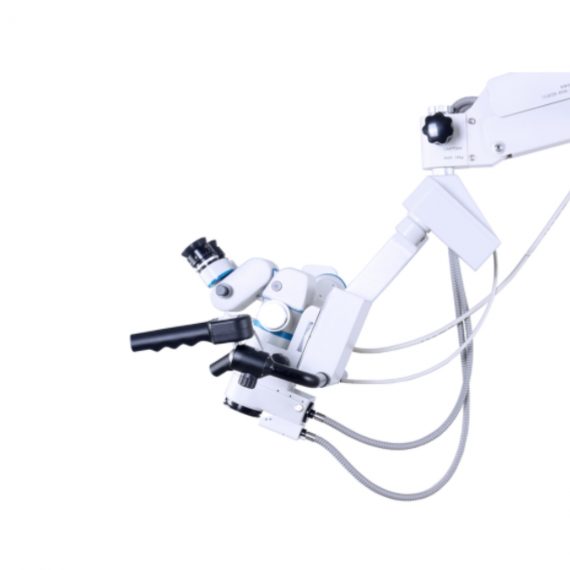
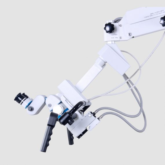


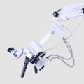
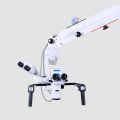
ENT/Neurosurgery Operating Microscope
$0.00
Shipped from abroad
Delivery & Availability:
Typically 14-21 working days – excluding furniture and heavy/bulky equipment. Please contact us for further information.
Corder Microscope has Fluid, Responsive and Accurate.
Fluid. Responsive. Accurate. These were a few of the principles guiding every phase in the design of the Corder Microscope. With the choicest mechanical machined components, the Corder Microscope has the grace and agility to adjust to every desired position on command. Well designed Apochromatic optics treated with Corder’s Mcoatings produce true-to life sharp images with high depth, definition and contrast.
Description
Features:
Corder Microscope has Fluid, Responsive and Accurate.
Fluid. Responsive. Accurate. These were a few of the principles guiding every phase in the design of the Corder Microscope. With the choicest mechanical machined components, the Corder Microscope has the grace and agility to adjust to every desired position on command. Well designed Apochromatic optics treated with Corder’s Mcoatings produce true-to life sharp images with high depth, definition and contrast.
More comfortable operation
Tiltable binocular tubes available, which can incline more than 60° depending on the posture and physique of the operating surgeon.
Movable range: 30° (straight) to 90° (inclined) Corder microscope configured with XYZ motorized movement operated through a comfortable foot /Handle control, a veryeffective co-axial illumnation and 50W halogen light source makes it ideal for Neuro surgeries.
Doctor-patient communication is easier
To address digital documentation needs, a host of digital SLR, video camera, and CCD adapters are made available with the ProLine in addition to Corder’s proprietary iVu multi-functional imaging solution. 1080P full hd image quality, efficient image management during the operation. Integrate your digital workflow to facilitate case management and facilitate more intuitive patient communication.
Technical Permeants:
Magnification: motorized zoom system, 1:6 zoom ratio, magnification 3x~16x
Focusing range: 50mm
Binocular tube: 30°~90° tiltable tube ,(0° ~200° optional)
Eyepiece: 12.5x / 10x
Objective lens: F 300mm(175mm, 250mm, 350mm optional)
pupil distance: 55mm~75mm
diopter adjustment: +6D ~ -6D
Field of view: Φ74~Φ12mm
X-Y translator: Motorized by foot switch or handle controller, ±30mm
Assistant tube: 360° Rotating assistant tube
Reset functions: YES
Illumination
System: Coaxial illumination
Light source: Halogen lamp
Light intensity adjustment: Continuous brightness adjustment 0-100000lux
Fiber optic illumination: Dual fiber
Field of illumination: Φ50mm
Filter: Red free filter, small spot
Accessories
CCD Camera system: Beam splitter, CCD adapter, CCD, Display
XENON LAMP: 150000lux
Integrated Video Adapter: SONY / CANON Camera
Click Here To Download Catalogue
Quick Comparison
| Settings | ENT/Neurosurgery Operating Microscope remove | DrGem Ceiling Analogue X-ray Machine remove | Bistos BT-770-12.1" Touchscreen Patient Monitor remove | DrGem Diamond All-In-One Digital X-ray Machine remove | ASPEL Ambulatory BP Machine remove | DrGem Floor Mounted Analogue X-ray remove |
|---|---|---|---|---|---|---|
| Name | ENT/Neurosurgery Operating Microscope remove | DrGem Ceiling Analogue X-ray Machine remove | Bistos BT-770-12.1" Touchscreen Patient Monitor remove | DrGem Diamond All-In-One Digital X-ray Machine remove | ASPEL Ambulatory BP Machine remove | DrGem Floor Mounted Analogue X-ray remove |
| Image | 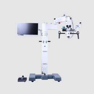 |  | 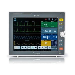 |  | 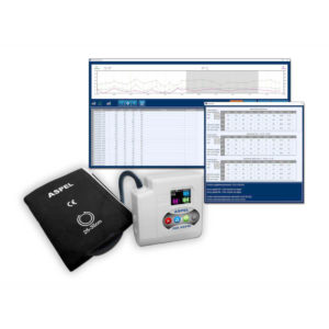 |  |
| SKU | SF1033560109-1 | SF1033560074-7 | SF1033560059-1 | SF1033560074-3 | SF1033560075-13 | SF1033560074-6 |
| Rating | ||||||
| Price |
|
| $902.00 |
| $920.00 |
|
| Stock | ||||||
| Availability | ||||||
| Add to cart | ||||||
| Description | Shipped from abroad
Corder Microscope has Fluid, Responsive and Accurate.Fluid. Responsive. Accurate. These were a few of the principles guiding every phase in the design of the Corder Microscope. With the choicest mechanical machined components, the Corder Microscope has the grace and agility to adjust to every desired position on command. Well designed Apochromatic optics treated with Corder's Mcoatings produce true-to life sharp images with high depth, definition and contrast. | Shipped from abroad The DrGem Ceiling Analogue X-ray Machine is a diagnostic radiography system that provides reliable high quality radiographic images with a reduced dose. The reliable high-frequency x-ray generators that are known worldwide for their excellent performance, lifetime and stability. Patient tables and wall stands are also offered. Delivery & Availability: Typically 21 working days – excluding furniture and heavy/bulky equipment. Please contact us for further information. | Shipped from Abroad The Bistos BT-770 patient monitor is equipped with a 12.1" touchscreen display, which allows for an easy operation and readability with a powerful rechargeable battery guaranteeing a continuous operation of 5 hours to monitor ECG, SpO2, NIBP, temperature and respiration Delivery & Availability: Typically 14 working days – excluding furniture and heavy/bulky equipment. Please contact us for further information. | Shipped from Abroad DrGem Diamond All-In-One Digital X-ray Machine is a fully automatic digital radiography system providing state-of-the-art image quality, image processing and user interface. With a wide selection of anatomical studies on the imaging software, DIAMOND automatically sets up the x-ray generator’s preprogrammed exposure technique settings, motorized radiographic stand positioning, x-ray collimation and post-image processing for the selected study. Specifically designed to increase workflow, this fully digital system offers convenient auto-positioning and advanced image processing to achieve big performance with little effort. Delivery & Availability: Typically 21 working days – excluding furniture and heavy/bulky equipment. Please contact us for further information. | Shipped from Abroad ASPEL Ambulatory BP Machine - is a recorder of long-term records of non-invasive measurement of blood pressure intended for use in clinics, hospitals, outpatient centers and specialist surgeries. The recorder enables the assessment of blood pressure by the oscillometric method in adult patients, pregnant women, including preeclampsia and pediatric patients (from 3 years of age). Blood pressure is assessed by using an inflatable cuff, an accurate pressure transducer, and a deflation valve. Delivery & Availability: Typically 10 working days – excluding furniture and heavy/bulky equipment. Please contact us for further information. | In Stock GXR Analogue X-ray system matches with a radiographic room which perfectly fits your workow and can be easily upgraded to DR system with the help of DR interface and PC interface in GXR generator as well as Bucky suitable to Flat Panel Detector. GXR X-ray system is equipped with a high frequency X-ray generator which consistently produces high quality radiograph in favor of high quality X-ray output with a very small kV ripple and accurate mA and mAs. GXR X-ray system is designed to provide convenience to operator and comfort to patient. Delivery & Availability: Typically 21 working days – excluding furniture and heavy/bulky equipment. Please contact us for further information. |
| Content | Features:Corder Microscope has Fluid, Responsive and Accurate.Fluid. Responsive. Accurate. These were a few of the principles guiding every phase in the design of the Corder Microscope. With the choicest mechanical machined components, the Corder Microscope has the grace and agility to adjust to every desired position on command. Well designed Apochromatic optics treated with Corder's Mcoatings produce true-to life sharp images with high depth, definition and contrast. More comfortable operation Tiltable binocular tubes available, which can incline more than 60° depending on the posture and physique of the operating surgeon. Movable range: 30° (straight) to 90° (inclined) Corder microscope configured with XYZ motorized movement operated through a comfortable foot /Handle control, a veryeffective co-axial illumnation and 50W halogen light source makes it ideal for Neuro surgeries.Doctor-patient communication is easierTo address digital documentation needs, a host of digital SLR, video camera, and CCD adapters are made available with the ProLine in addition to Corder's proprietary iVu multi-functional imaging solution. 1080P full hd image quality, efficient image management during the operation. Integrate your digital workflow to facilitate case management and facilitate more intuitive patient communication. Technical Permeants: Magnification: motorized zoom system, 1:6 zoom ratio, magnification 3x~16x Focusing range: 50mm Binocular tube: 30°~90° tiltable tube ,(0° ~200° optional) Eyepiece: 12.5x / 10x Objective lens: F 300mm(175mm, 250mm, 350mm optional) pupil distance: 55mm~75mm diopter adjustment: +6D ~ -6D Field of view: Φ74~Φ12mm X-Y translator: Motorized by foot switch or handle controller, ±30mm Assistant tube: 360° Rotating assistant tube Reset functions: YES Illumination System: Coaxial illumination Light source: Halogen lamp Light intensity adjustment: Continuous brightness adjustment 0-100000lux Fiber optic illumination: Dual fiber Field of illumination: Φ50mm Filter: Red free filter, small spot Accessories CCD Camera system: Beam splitter, CCD adapter, CCD, Display XENON LAMP: 150000lux Integrated Video Adapter: SONY / CANON CameraClick Here To Download Catalogue | DrGem Ceiling Analogue X-ray Machine is a diagnostic radiography system X-ray Machine that provides reliable high quality radiographic images with a reduced dose. The reliable high-frequency x-ray generators that are known worldwide for their excellent performance, lifetime and stability. Patient tables and wall stands are also offered.
Features of DrGem Ceiling Analogue X-ray Machine
Click Here To Download Catalogue |
Bistos BT-770 is a 12.1" touchscreen patient monitor designed for easy operations.
SPECIFICATIONS
Click Here To Download Catalogue | DrGem Diamond All-In-One Digital X-ray Machine is a fully automatic digital radiography system providing state-of-the-art image quality, image processing and user interface. With a wide selection of anatomical studies on the imaging software, DIAMOND automatically sets up the x-ray generator’s pre-programmed exposure technique settings, motorized radiographic stand positioning, x-ray collimation and post-image processing for the selected study. Specifically designed to increase workflow, this fully digital system offers convenient auto-positioning and advanced image processing to achieve big performance with little effort.
Features of DrGem Diamond All-In-One Digital X-ray Machine:
Outstanding Image Quality -
Digital radiography via at panel detector improves your workflow, exam speed and comfort with efficiency. Digital at panel detector with Csl screen provides excellent spatial resolution, MTF, DQE and stability based on ne pixel pitch. A 3-field ion-chamber is provided for AEC function.
Automatic Collimation –
Automatic x-ray eld size control of the motorized collimator corresponds to dierent SIDs. Includes user adjustable lamp timer with on/oswitch.
Automatic Positioning –
Click Here To Download Catalogue | ASPEL Ambulatory BP Machine - is a recorder of long-term records of non-invasive measurement of blood pressure intended for use in clinics, hospitals, outpatient centers and specialist surgeries. The recorder enables the assessment of blood pressure by the oscillometric method in adult patients, pregnant women, including preeclampsia and pediatric patients (from 3 years of age). Blood pressure is assessed by using an inflatable cuff, an accurate pressure transducer, and a deflation valve.
Features:
Save-2-Safe: Double security system
Thanks to the use of two independent measuring systems with an additional valve, it meets the highest standards and takes care of patient safety even better.
Start-Easy: Quick start in two moves
The quick launch function allows you to use the device instantly, easily allows you to start recording in holter mode.
Memo-Care: Cuff pressure memory
Recorder remembers the pressure in the cuff. Thanks to the use of Intelligent Solutions, it adapts individually to the patient.
Power-Usb: USB connection
The device can work without batteries: by connecting to a computer via a USB cable.
Technical Specification:
Click Here To Download Catalogue | DrGem GXR Floor Mounted Analogue X-ray system matches with a radiographic room which perfectly fits your workflow and can be easily upgraded to DR system with the help of DR interface and PC interface in GXR generator as well as Bucky suitable to Flat Panel Detector. GXR (Analogue X-ray)system is equipped with a high frequency X-ray generator which consistently produces high quality radiograph in favor of high quality X-ray output with a very small kV ripple and accurate mA and mAs. GXR (Analogue X-ray) system is designed to provide convenience to operator and comfort to patient.
Features of DrGem GXR Floor Mounted Analogue X-ray:
Click Here To Download Catalogue |
| Weight | N/A | N/A | N/A | N/A | N/A | N/A |
| Dimensions | N/A | N/A | N/A | N/A | N/A | N/A |
| Additional information |


Reviews
There are no reviews yet.